. 2021 Oct 21;10(11):1654.
doi: 10.3390/antiox10111654.

Neuroprotective Activity of Melittin-The Main Component of Bee Venom-Against Oxidative Stress Induced by Aβ25-35 in In Vitro and In Vivo Models

Affiliations

Neuroprotective Activity of Melittin-The Main Component of Bee Venom-Against Oxidative Stress Induced by Aβ25-35 in In Vitro and In Vivo Models

Cong Duc Nguyen et al. Antioxidants (Basel). .

Abstract

Melittin, a 26-amino acid peptide, is the main component of the venom of four honeybee species and exhibits neuroprotective actions. However, it is unclear how melittin ameliorates neuronal cells in oxidative stress and how it affects memory impairment in an in vivo model. We evaluated the neuroprotective effect of melittin on Aβ25-35-induced neuro-oxidative stress in both in vitro HT22 cells and in vivo animal model. Melittin effectively protected against HT22 cell viability and significantly deregulated the Aβ25-35-induced overproduction of intracellular reactive oxygen species. Western blot analysis showed that melittin suppressed cell apoptosis and regulated Bax/Bcl-2 ratio, as well as the expression of proapoptotic related factors: Apoptosis-inducing factor (AIF), Calpain, Cytochrome c (CytoC), Cleaved caspase-3 (Cleacas3). Additionally, melittin enhanced the antioxidant defense pathway by regulating the nuclear translocation of nuclear factor erythroid 2-like 2 (Nrf2) thus upregulated the production of the heme oxygenase-1 (HO-1), a major cellular antioxidant enzyme combating neuronal oxidative stress. Furthermore, melittin treatment activated the Tropomyosin-related kinase receptor B (TrkB)/cAMP Response Element-Binding (CREB)/Brain-derived neurotrophic factor (BDNF), contributing to neuronal neurogenesis, and regulating the normal function of synapses in the brain. In our in vivo experiment, melittin was shown to enhance the depleted learning and memory ability, a novel finding. A mouse model with cognitive deficits induced by Aβ25-35 intracerebroventricular injection was used. Melittin had dose-dependently enhanced neural-disrupted animal behavior and enhanced neurogenesis in the dentate gyrus hippocampal region. Further analysis of mouse brain tissue and serum confirmed that melittin enhanced oxidant-antioxidant balance, cholinergic system activity, and intercellular neurotrophic factors regulation, which were all negatively altered by Aβ25-35. Our study shows that melittin exerts antioxidant and neuroprotective actions against neural oxidative stress. Melittin can be a potential therapeutic agent for neurodegenerative disorders.

Keywords: BDNF; bee venom; beta amyloid; melittin; neurodegeneration; oxidative stress.


Conflict of interest statement

The authors declare no conflict of interest.

Figures

Figure 1
Figure 1
Screening for melittin maximum safety dosage and melittin protective effect against Aβ25–35 stress induced in HT22 cells. (A) Screening for maximum melittin safety concentration. The result indicated that 3 μM was the highest safety concentration of melittin to be studied. To conduct this experiment: Cells were seeded in 96-well plates; after incubation for 24 h, Melittin at different concentrations was introduced, and cell availability was examined after 24 h of incubation. (B) Protective effect of melittin against Aβ25–35 stress. The result indicated that 0.3 to 3 μM melittin was found to dosage-dependently ameliorate HT22 cell availability. To conduct this experiment: Cells were seeded in 96-well plates; after incubation for 24 h, melittin from 0.1 to 3 μM was introduced 1 h before Aβ25–35 (7 μM) challenge, and cell availability was examined after 24 h of incubation. Data are presented as the mean ± standard deviation values of triple determinations. # p < 0.01 vs. control * p < 0.05 and ** p < 0.01 vs. Aβ25–35 only-treated group.
Figure 2
Figure 2
Apoptosis-inhibitory effects of melittin on Aβ25–35 induced HT-22 cells. (A) Immunofluorescence analysis to determine the cellular apoptosis rate. The result indicated that melittin at 0.3 to 3 μM dosage-dependently ameliorated HT22 cell apoptosis under Aβ25–35 (7 μM) stress-induced conditions. To conduct this experiment: Seven hours after Aβ25–35 (7 μM) challenge in 96-well plates, immunofluorescence staining was carried out to exhibit live cells (blue, stained by CytoCalcein Violet 450), necrotic cells (red, indicated by 7-AAD staining), and apoptotic cells (green, Apopxin Green Indicator). (B) Western blot analysis of key apoptosis proteins. The result indicated that melittin at 0.3 to 3 μM dosage-dependently normalized the expression of pro and anti-apoptosis protein under Aβ25–35 stress challenge. To conduct this experiment: Seven hours after Aβ25–35 (7 μM) challenge in 6-well plates, cells were lysed, and western blot was performed. Data are presented as mean ± standard deviation values of triple determinations. # p < 0.01 vs. control * p < 0.05 and ** p < 0.01 vs. Aβ25–35 only-treated group.
Figure 3
Figure 3
Melittin regulates cellular oxidative stress induced by Aβ25–35 in HT22 cells. (A) Immunofluorescence analysis to determine the cellular ROS rate. The result indicated that melittin at 0.3 to 3 μM dosage-dependently normalized the expression of pro and anti-apoptosis proteins under Aβ25–35 stress challenge. To conduct this experiment: Seven hours after Aβ25–35 (7 μM) challenge in 96-well plates, immunofluorescence analysis by DCFDA staining was carried out. (B) Cellular MDA, LDH, and protein carbonyls levels. The result indicated that melittin at 0.3 to 3 μM dosage-dependently down-regulated MDA, LDH, and protein carbonyl parameters under Aβ25–35 stress challenge. To conduct this experiment: Seven hours after Aβ25–35 (7 μM) challenge in 6-well plates, kits measuring MDA, LDH, and protein carbonyls were used to determine the parameters. Data are presented as mean ± standard deviation values of triple determinations. # p < 0.01 vs. control * p < 0.05 and ** p < 0.01 vs. Aβ25–35 only-treated group.
Figure 4
Figure 4
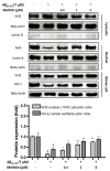
Effect of melittin on nuclear translocation of Nrf2 and an increase in the production of the antioxidant enzyme HO-1. The result suggested that melittin at 0.3 to 3 μM dosage-dependently enhanced Nrf2 nuclear translocation and as a result increased the expression of the HO-1 antioxidant enzyme in HT22 cells under Aβ25–35 (7 μM) challenge. To conduct this experiment: Seven hours after Aβ25–35 (7 μM) challenge in 6-well plates, whole cell, cytosolic, and nuclear proteins were obtained, and western blot was conducted. The membranes of Nrf2, Lamin B, and Beta actin in cytosolic extract and the membranes of their respective counterparts in nuclear extract under similar treatments and imaging exposure time to express the true scale of protein expression between nuclear and cytosolic proteins. Total Lamin B signal of the cytosolic extract was less than 5% of that in the nuclear extract, total Beta actin in the cytosolic extract was less than 5% of that in the nuclear extract (numeric bars not shown); this indicates the reliability of the manual extraction conducted. Data are presented as mean ± standard deviation values of triple determinations. # p < 0.01 vs. Control * p < 0.05 and ** p < 0.01 vs. Aβ25–35 only-treated group.
Figure 5
Figure 5
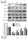
Effects of melittin on the activation of brain-derived neurotrophic factor signaling. The result suggested that melittin enhanced the performance of the BDNF/TrkB/CREB pathway under Aβ25–35 (7 μM) challenge. To conduct this experiment: Seven hours after Aβ25–35 (7 μM) challenge, cells were lysed, and western blot was performed. Data are presented as mean ± standard deviation values of triple determinations. # p < 0.01, vs. control * p < 0.05, ** p < 0.01, vs. Aβ25–35 only-treated group.
Figure 6
Figure 6
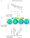
Protective effect of melittin on memory in Aβ25–35-treated mice studied in a Morris water maze experiment. Two tests (A,B) exhibited a decrease in learning and memory performance when mice were induced with Aβ25–35 ICV., and melittin 1.5 mg/kg significantly reversed this cognitive disfunction effect. The water maze behavior experiment recorded the escape latency time on days 7–10. (A) Animal escape latency performance on days 7 to 10. To conduct this experiment: Mice are administrated melittin (1.5 or 0.15 mg/kg SC.) after Aβ25–35 treatment (5 μg/mouse, ICV.), a training session was conducted on day 6, and the escape latency test was performed on days 7 to 10. (B) A probe test was carried out at the end of day 10 to further reinforce behavior evaluations. To conduct this experiment: The platform was removed, and mice were let to swim freely for 2 min to determine their memory of the disappeared platform location. The color scale indicates the average distribution position of animals within each group. The circle located in the upper-left quadrant represents the platform location. Data are presented as mean ± standard error values of the sextuple determinations. # p < 0.01 vs. control ** p < 0.01 vs. Aβ25–35 only-treated group.
Figure 7
Figure 7
Protective effect of melittin on neuronal cells in the dentate gyrus region against Aβ25–35 induced stress. The result indicated that Aβ25–35 ICV. treatment reduced neuron cell neurogenesis significantly, and this was reversed by melittin. To conduct this experiment: Brain sections stained with doublecortin antibody, and the hippocampal dentate gyrus area were examined. Data are presented as the mean ± standard error of the sextuple determinations. # p < 0.01 vs. control * p < 0.05 and ** p < 0.01 vs. Aβ25–35 only-treated group.
Figure 8
Figure 8
Major antioxidant parameters were examined in in vivo samples. The result showed that Aβ25–35 ICV. treatment significantly increased oxidative stress in the hippocampus and serum, which was reversed by melittin treatment. To conduct this experiment: Hippocampus tissue and serum were prepared, and ROS, NO, and MDA levels were examined as described in the materials and methods section. Data are presented as the mean ± standard error of the sextuple determinations. # p < 0.01 vs. control ** p < 0.01 vs. Aβ25–35 only treated group.
Figure 9
Figure 9
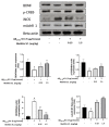
Levels of BDNF, p-CREB, iNOS, and mAchR1 protein expression in the mouse hippocampus show the dose-dependent protective effect of melittin against Aβ25–35-induced neuro-imbalance in vivo. To conduct this experiment: Western blot experiment was performed on protein extracts obtained from hippocampus tissue as described in the materials and methods section. Data are presented as the mean ± standard error values of the sextuple determinations. # p < 0.01 vs. control * p < 0.05 and ** p < 0.01 vs. Aβ25–35 only-treated group.
Figure 10
Figure 10
Levels of AchE activity and Ach content in the mouse brain. The cholinergic system exhibited ameliorations as melittin was administrated, against Aβ25–35 ICV.-induced neurodegeneration. To conduct this experiment: Analysis were performed on hippocampal tissue as described in the materials and methods section. Data are presented as the mean ± standard error of the sextuple determinations. # p < 0.01 vs. control * p < 0.05 and ** p < 0.01 vs. Aβ25–35 only-treated group.

Similar articles

Cited by

References

    1. Hwang D.S., Kim S.K., Bae H. Therapeutic effects of bee venom on immunological and neurological diseases. Toxins. 2015;7:2413–2421. doi: 10.3390/toxins7072413. - DOI - PMC - PubMed
    1. Frangieh J., Salma Y., Haddad K., Mattei C., Legros C., Fajloun Z., Obeid D. El First characterization of the venom from apis mellifera syriaca, a honeybee from the middle east region. Toxins. 2019;11:191. doi: 10.3390/toxins11040191. - DOI - PMC - PubMed
    1. Somwongin S., Chantawannakul P., Chaiyana W. Antioxidant activity and irritation property of venoms from Apis species. Toxicon. 2018;145:32–39. doi: 10.1016/j.toxicon.2018.02.049. - DOI - PubMed
    1. Moon D.O., Park S.Y., Lee K.J., Heo M.S., Kim K.C., Kim M.O., Lee J.D., Choi Y.H., Kim G.Y. Bee venom and melittin reduce proinflammatory mediators in lipopolysaccharide-stimulated BV2 microglia. Int. Immunopharmacol. 2007;7:1092–1101. doi: 10.1016/j.intimp.2007.04.005. - DOI - PubMed
    1. Han S.M., Kim J.M., Park K.K., Chang Y.C., Pak S.C. Neuroprotective effects of melittin on hydrogen peroxide-induced apoptotic cell death in neuroblastoma SH-SY5Y cells. BMC Complement. Altern. Med. 2014;14:1–8. doi: 10.1186/1472-6882-14-286. - DOI - PMC - PubMed