. 2021 Oct 28;10(11):1717.
doi: 10.3390/antiox10111717.

Peucedanum japonicum Thunberg and Its Active Components Mitigate Oxidative Stress, Inflammation and Apoptosis after Urban Particulate Matter-Induced Ocular Surface Damage

Affiliations

Peucedanum japonicum Thunberg and Its Active Components Mitigate Oxidative Stress, Inflammation and Apoptosis after Urban Particulate Matter-Induced Ocular Surface Damage

Wan Seok Kang et al. Antioxidants (Basel). .

Abstract

We previously demonstrated that urban particulate matter (UPM) exposure decreases the migration activity and survival of human corneal epithelial cells (HCECs). Herein, we investigated the potential to improve the corneal wound-healing ability of Peucedanum japonicum Thunb. leaf extract (PJE) and its active components on UPM-induced ocular surface damage in vitro and in vivo. PJE effectively assisted wound healing without altering HCEC survival and enhanced catalase (CAT), heme oxygenase 1 (HO1) and glutathione peroxidase 1 (GPX1) antioxidant gene expression. A corneal wound was uniformly induced on the right eye in all experimental animals and divided into eight groups such as two control groups (wounded right eye group-NR and non-wounded left eye group-NL), UPM treated group and PJEs (25, 50, 100, 200, 400 mg/kg) treated groups. Corneal abrasion model rats exposed to UPM showed delayed wound healing compared to unexposed rats, but wound healing was dose-dependently enhanced by PJE oral administration. Seventy-two hours after wound generation, inflammatory cells, apoptotic cells and interleukin-6 (IL-6) expression were increased substantially after UPM exposure, but PJE treatment significantly reduced the wound to an almost normal level while enhancing re-epithelialization without changing corneal thickness. Next, we tried to identify the key molecules for enhancing wound healing through fractionation. The major compounds in the fraction, confirmed by high-performance liquid chromatography (HPLC), were chlorogenic acid (CA), neochlorogenic acid (NCA) and cryptochlorogenic acid (CCA). Each type of CA isomers showed slightly different half maximal effective (EC50) and maximal effective (ECmax) concentrations, and their mixtures synergistically enhanced HCEC migration. Thus, corneal abrasion wound recovery after UPM exposure improved after PJE treatment, and the active PJE components were identified, providing an important basis to develop therapeutics for ocular surface damage using PJE.

Keywords: Peucedanum japonicum Thunberg; antioxidant; corneal epithelial cells; inflammation; urban particulate matter (UPM); wound healing.


Conflict of interest statement

The authors declare no conflict of interest.

Figures
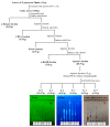
Figure 1
Figure 1
Scheme of PJE fractionation by solvent-solvent extraction and open column chromatography. TLC analysis images are also presented.
Figure 2
Figure 2
HPLC chromatograms of chlorogenic acid (CA), neochlorogenic acid (NCA) and cryptochlorogenic acid (CCA) as standard compounds and the P. japonicum Thunberg leaf extract (PJE).
Figure 3
Figure 3
Effects of UPM and PJEs on the migration activity were assessed on the human corneal epithelial cells (HCECs). (A) A scratch was induced to the HCECs treated with UPM and various concentrations of PJE for 24 h. The closed area after 24 h from scratch area were calculated. The scale bars indicate 400 μm. (B) The relative migration rates of HCECs are expressed as the means ± SD. (C) The survival rates of HCECs are expressed as the means ± SD. ### p < 0.001 compared to Con; * p < 0.05, ** p < 0.05, *** p < 0.001 compared to 0 μg/mL PJE; ns, not significant.
Figure 4
Figure 4
Effects of PJE on the mRNA expression of antioxidative genes in HCECs. After the scratch assay, the relative mRNA expression levels of (A) SOD1, (B) CAT, (C) HO1 and (D) GPX1 were analyzed, and the fold changes are presented compared with the Con as the means ± SD. # p < 0.05, ## p < 0.01 compared to Con; * p < 0.05, ** p < 0.01, *** p< 0.001 compared to 0 μg/mL PJE; ns, not significant.
Figure 5
Figure 5
Effects of PJE in a corneal abrasion model after UPM exposure. PJE and UPM were used as pretreatments for 5 days, and 4 mm wounds were generated on the right cornea. Images were obtained at 0, 8, 16, 24, 36, 48 and 72 h by fluorescein staining. (A) Time course images of corneal wound healing in the normal right eye (NR, normal wound), UPM and 400 mg/kg PJE groups. (B) Representative images of each group at 36 h. (C) The wound healing areas were calculated and are presented as the means ± SD. ## p < 0.01 compared to NR; * p < 0.05, ** p < 0.01, *** p < 0.001 compared to UPM.
Figure 6
Figure 6
Histological changes during corneal wound healing after treatment with PJE and after UPM exposure. (A) Images of hematoxylin and eosin (H&E) staining of the cornea at 72 h were acquired at 200× magnification, and representative images are shown. Red arrow, re-epithelialized epithelium; black arrow, cell attachment on the endothelium. The infiltration of immune cells in (B) the central stroma and (C) the lateral stroma, (D) the re-epithelialization rate and (E) cell attachment to the endothelium were calculated, and the results are presented as the means ± SD.  p < 0.05, †† p < 0.01 compared to NL; ### p < 0.001 compared to NR; ** p < 0.01, *** p < 0.001 compared to UPM.
Figure 7
Figure 7
Immunohistological changes in IL-6 expression during corneal wound healing after treatment with PJE and after UPM exposure. (A) Immunohistochemical staining images for IL-6 in the corneas after 72 h were acquired at 200× magnification, and representative images are shown. (B) The intensities of the stained areas were calculated and the data are presented as the means ± SD. (C) The corneal thicknesses were calculated and are presented as the means ± SD.  p < 0.05 compared to NL; ## p < 0.01 compared to NR; * p < 0.05, ** p < 0.01 compared to UPM; ns, not significant.
Figure 8
Figure 8
Apoptotic cells during corneal wound healing after treatment with PJE and after UPM exposure. (A) TUNEL staining images of the cornea at 72 h were acquired at 200× magnification, and representative images are shown. (B) The intensities of the apoptotic cells were calculated, and the results are presented as the means ± SD.  p < 0.05 compared to NL; ### p < 0.001 compared to NR. The p values of all PJE groups were below 0.001 compared to UPM.
Figure 9
Figure 9
Fractions of PJE after solvent fractionations and their effects on HCEC wound healing. (A) HPLC chromatograms of the PJE and its fractions. (B) The migration rates of each fraction were calculated, and data from the concentrations of 1 μg/mL and 3 μg/mL are presented as the means ± SD. (C) The migration rates of the (C) BuOH fraction and (D) water fraction are presented as the means ± SD. ††† p < 0.001 compared to Con; * p < 0.05, ** p < 0.01 compared to 0 μg/mL PJE.
Figure 10
Figure 10
HP-20 open column chromatography of the water fraction of PJE (PJE/W) and the effects of these fractions on HCEC wound healing. (A) HPLC chromatograms of PJE and its fractions (F0~F5). (B) The migration rates of each fraction were calculated, and data from the concentrations of 1 μg/mL and 3 μg/mL are presented as the means ± SD. (C) The migration rates of (C) F1 and (D) F5 are presented as the means ± SD. ††† p < 0.001 compared to Con; * p < 0.05, ** p < 0.01 *** p < 0.001 compared to 0 μg/mL PJE.
Figure 11
Figure 11
Effects of the major components of PJE on wound healing. (A) The migration rates of each CA and their mixtures were calculated, and data from the concentrations of 0.846 μM and 8.46 μM are presented as the means ± SD. The relative migration rates of (B) each CA individually, (C) double CA mixtures and (D) the triple CA mixture are presented as the means ± SD; their EC50 values are indicated by dotted lines. ††† p < 0.001 compared to Con; * p < 0.05, ** p < 0.01 *** p < 0.001 compared to 0 μg/mL PJE.

References

    1. Valko M., Leibfritz D., Moncol J., Cronin M.T., Mazur M., Telser J. Free radicals and antioxidants in normal physiological functions and human disease. Int. J. Biochem. Cell Biol. 2007;39:44–84. doi: 10.1016/j.biocel.2006.07.001. - DOI - PubMed
    1. Guo Y., Jia Y., Pan X., Liu L., Wichmann H.-E. The association between fine particulate air pollution and hospital emergency room visits for cardiovascular diseases in Beijing, China. Sci. Total Environ. 2009;407:4826–4830. doi: 10.1016/j.scitotenv.2009.05.022. - DOI - PubMed
    1. Camara J.G., Lagunzad J.K.D. Ocular findings in volcanic fog induced conjunctivitis. Hawaii Med. J. 2011;70:262. - PMC - PubMed
    1. Tecer L.H., Alagha O., Karaca F., Tuncel G., Eldes N. Particulate matter (PM2.5, PM10-2.5, and PM10) and children’s hospital admissions for asthma and respiratory diseases: A bidirectional case-crossover study. J. Toxicol. Environ. Health Part A. 2008;71:512–520. doi: 10.1080/15287390801907459. - DOI - PubMed
    1. Shao Y. Air pollutant particulate matter 2.5 induces dry eye syndrome in mice. Investig. Ophthalmol. Vis. Sci. 2018;59:3839. - PMC - PubMed

LinkOut - more resources