. 2021 Nov 11;10(11):1798.
doi: 10.3390/antiox10111798.

Calcium Dobesilate Modulates PKCδ-NADPH Oxidase- MAPK-NF-κB Signaling Pathway to Reduce CD14, TLR4, and MMP9 Expression during Monocyte-to-Macrophage Differentiation: Potential Therapeutic Implications for Atherosclerosis

Affiliations

Calcium Dobesilate Modulates PKCδ-NADPH Oxidase- MAPK-NF-κB Signaling Pathway to Reduce CD14, TLR4, and MMP9 Expression during Monocyte-to-Macrophage Differentiation: Potential Therapeutic Implications for Atherosclerosis

Florence Njau et al. Antioxidants (Basel). .

Abstract

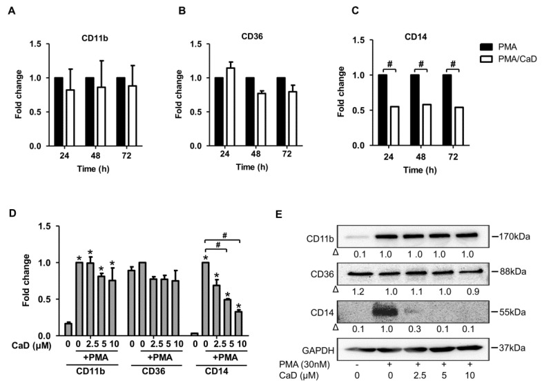
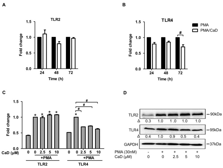
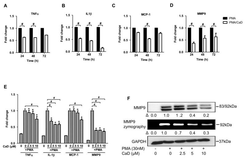
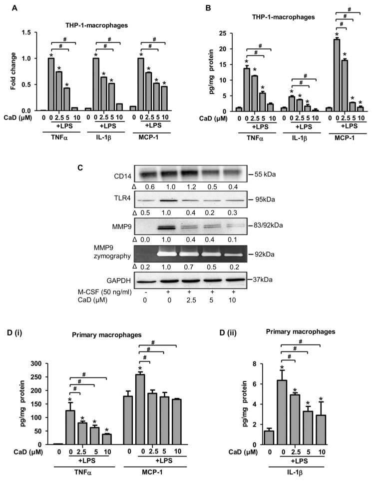
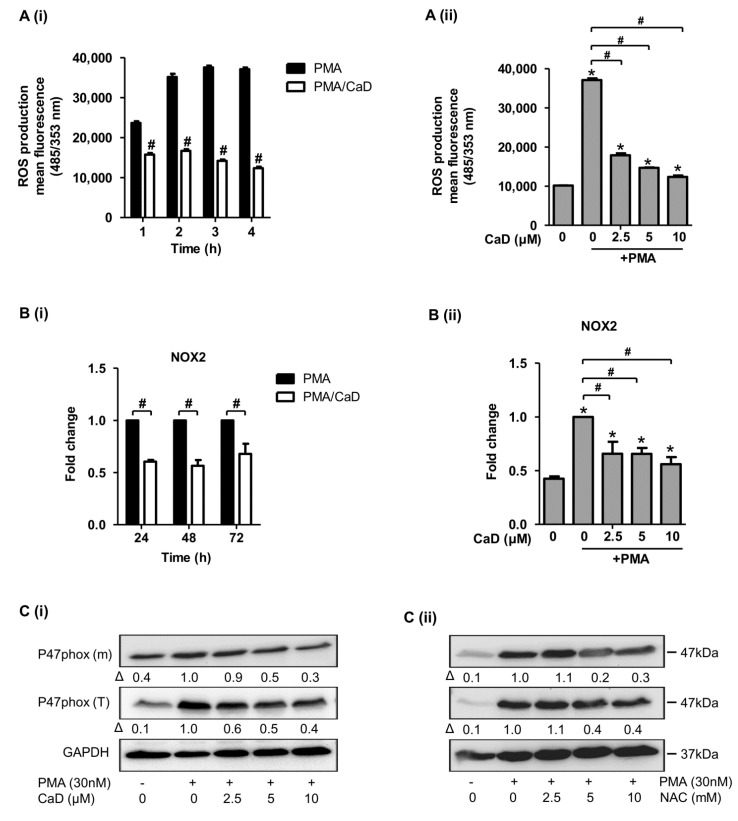
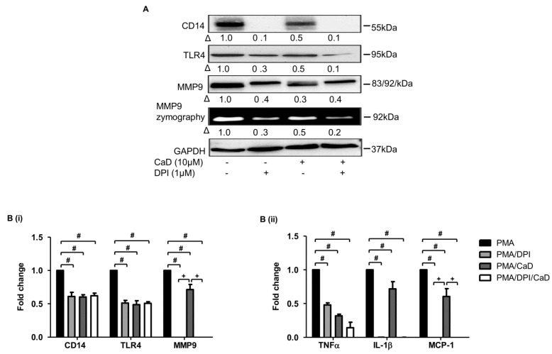
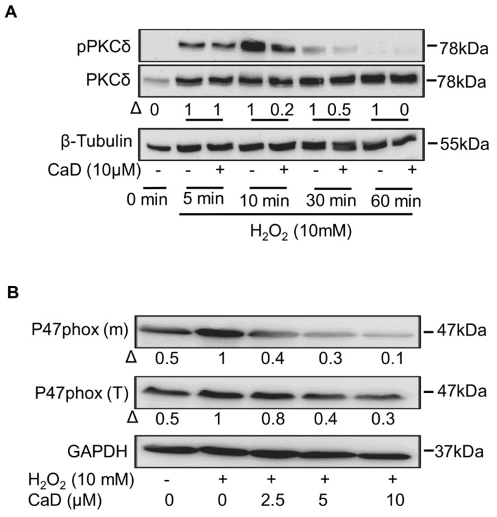
Monocyte-to-macrophage differentiation results in the secretion of various inflammatory mediators and oxidative stress molecules necessary for atherosclerosis pathogenesis. Consequently, this differentiation represents a potential clinical target in atherosclerosis. Calcium dobesilate (CaD), an established vasoactive and angioprotective drug in experimental models of diabetic microvascular complications reduces oxidative stress and inhibits inflammation via diverse molecular targets; however, its effect on monocytes/macrophages is poorly understood. In this study, we investigated the anti-inflammatory mechanism of CaD during phorbol 12-myristate 13-acetate (PMA)-induced monocyte-to-macrophage differentiation in in vitro models of sepsis (LPS) and hyperglycemia, using THP-1 monocytic cell line. CaD significantly suppressed CD14, TLR4, and MMP9 expression and activity, lowering pro-inflammatory mediators, such as IL1β, TNFα, and MCP-1. The effects of CaD translated through to studies on primary human macrophages. CaD inhibited reactive oxygen species (ROS) generation, PKCδ, MAPK (ERK1/2 and p38) phosphorylation, NOX2/p47phox expression, and membrane translocation. We used hydrogen peroxide (H2O2) to mimic oxidative stress, demonstrating that CaD suppressed PKCδ activation via its ROS-scavenging properties. Taken together, we demonstrate for the first time that CaD suppresses CD14, TLR4, MMP9, and signature pro-inflammatory cytokines, in human macrophages, via the downregulation of PKCδ/NADPH oxidase/ROS/MAPK/NF-κB-dependent signaling pathways. Our data present novel mechanisms of how CaD alleviates metabolic and infectious inflammation.

Keywords: PKCδ; atherosclerosis; calcium dobesilate; inflammation; monocyte-macrophage differentiation; oxidative stress.


Conflict of interest statement

The authors declare no conflict of interest.

Figures

Figure 1
Figure 1
Calcium dobesilate (CaD) inhibits PMA-induced CD14 expression. THP-1 monocytes were pretreated with 10 µmol/L CaD for 1 h, followed by stimulation with PMA (30 nmol/L) for various time points (AC), or treated with various concentrations (0–10 µmol/L) of CaD for 1 h (D,E) followed by PMA treatment for 72 h. Transcript levels of the indicated genes were measured by quantitative RT-PCR (AD), and protein levels measured by Western blotting (E). Δ, fold-change normalized to PMA only (n = 3–4, mean ± SEM. * p < 0.05 vs. no treatment, # p < 0.05 vs. PMA only, Student’s t-test (AC), or one-way ANOVA (D)). The Western blot represents one from at least three independent experiments.
Figure 2
Figure 2
CaD on TLR4 expression during monocyte-to-macrophage differentiation. THP-1 monocytes were pretreated with 10 µmol/L of CaD for 1 h followed by stimulation with PMA (30 nmol/L) for various time points (A,B), or THP-1 monocytes were treated with various concentrations (0–10 µmol/L) of CaD for 1 h (CD) followed by PMA treatment for 72 h. TLR2 and TLR4 transcripts (AC) and protein levels (D) were measured by quantitative RT-PCR and Western blotting, respectively. Δ, fold-change normalized to PMA only (n = 3–4, mean ± SEM. * p < 0.05 vs. no treatment, # p < 0.05 vs. PMA only, Student’s t-test (A,B), or one-way ANOVA (C)). The Western blot represents one from at least three independent experiments.
Figure 3
Figure 3
CaD inhibits PMA-induced inflammation during PMA-induced monocyte-to-macrophage differentiation. THP-1 monocytes were pretreated with CaD (10 µmol/L) for 1 h followed by stimulation with PMA (30 nmol/L) for various time points (AD), or THP-1 monocytes were treated with various CaD concentrations (0–10 µmol/L), followed by PMA treatment for 48 h (E,F). TNFα, IL-1β, MCP-1, and MMP9 transcript levels were measured by quantitative RT-PCR, and MMP9 protein levels and activity were measured by Western blotting and gelatin zymography, respectively. Δ, fold-change normalized to PMA only (n = 3–4, mean ± SEM. * p < 0.05 vs. no treatment, # p < 0.05 vs. PMA only, Student’s t-test (AD), or one-way ANOVA (E)). The Western blots represent one from at least three independent experiments.
Figure 4
Figure 4
CaD inhibits primary human monocyte-to-macrophage differentiation and LPS-induced inflammation. Primary human monocytes were treated with various CaD concentrations (0–10 µmol/L) for 1 h followed by M-CSF (50 ng/mL) treatment for 5 days (A). Protein levels and MMP9 activity were measured by Western blotting and gelatin zymography, respectively. THP-1 cells and primary human monocytes were differentiated into macrophages in the presence of various CaD concentrations (0–10 µmol/L) for 48 h and 5 days, respectively (BD). Macrophages were then stimulated with LPS (100 ng/mL) for 24 h. TNFα, IL1β, and MCP-1 transcript levels (B) were measured by quantitative RT-PCR, and protein levels (C,D) in the conditioned media were measured by ELISA (B). Δ, fold-change normalized to PMA only (n = 3, mean ± SEM. * p < 0.05 vs. no LPS, # p < 0.05 vs. LPS only, one-way ANOVA).
Figure 5
Figure 5
Effect of CaD on PKCδ-MAPK signaling during PMA-induced monocyte-to-macrophage differentiation. THP-1 monocytes were pretreated with various concentrations (0–10 µmol/L) of CaD for 1 h, followed by PMA treatment for 24 h (A) or 30 min (B). PKCα, PKCßII, and PKCδ transcript levels (A) were measured by quantitative RT-PCR, and phosphorylation of PKC and MAPK pathway components (B) was measured by Western blotting. Δ, fold-change normalized to PMA only (n = 3, mean ± SEM. * p < 0.05 vs. no treatment, # p < 0.05 vs. PMA only, one-way ANOVA). The Western blots represent one from at least three independent experiments.
Figure 6
Figure 6
Inhibition of monocyte-to-macrophage differentiation and inflammation by CaD involves PKCδ-dependent signaling. THP-1 monocytes were pretreated with the PKCδ inhibitor rottlerin (Rot) for 1 h, followed by CaD for 1 h, and then treated with PMA (30 nmol/L) for 72 h. The differentiation and inflammation marker expression were measured by Western blotting (A), MMP9 activity measured by gelatin zymography, and transcript levels (B) measured by quantitative RT-PCR. Δ, fold-change normalized to PMA only (n = 3, mean ± SEM. # p < 0.05 vs. PMA only, one-way ANOVA). The Western blot represents one from at least three independent experiments.
Figure 7
Figure 7
CaD suppresses the MAPK signaling to abolish monocyte-to-macrophage differentiation and inflammation. THP-1 monocytes were pretreated with an ERK1/2 (U0126) or P38 inhibitor (SB203580) for 1 h, followed by CaD for 1 h, and then treated with PMA (30 nmol/L) for 72 h. The differentiation and inflammation markers expression were measured by Western blotting (A), MMP9 activity was measured by gelatin zymography, and transcript levels (B,C) were measured by quantitative RT-PCR. Δ, fold-change, normalized to PMA only (n = 3, mean ± SEM. + p < 0.05 vs. inhibitor treatment, # p < 0.05 vs. PMA only, one-way ANOVA). The Western blots represent one from at least three independent experiments.
Figure 8
Figure 8
CaD inhibits monocyte-to-macrophage differentiation and inflammation in an NF-κB-dependent manner. THP-1 monocytes were pretreated with various concentrations (0–10 µmol/L) of CaD for 1 h, followed by PMA treatment for 10 min (A). THP-1 monocytes were pretreated with an NF-κB inhibitor (Bay 11-7085) for 1 h, followed by CaD for 1 h, and then treated with PMA (30 nmol/L) for 72 h (B,C). IKBα phosphorylation and the differentiation and inflammation marker expression (A,B) were measured by Western blotting, MMP9 activity was measured by gelatin zymography, and transcript (C) levels were measured by quantitative RT-PCR. Δ, fold change normalized to PMA only (n = 3, mean ± SEM. + p < 0.05 vs. Bay treatment, # p < 0.05 vs. PMA only, one-way ANOVA). The Western blots represent one from at least three independent experiments.
Figure 9
Figure 9
CaD modulates the ROS-NADPH oxidase pathway during monocyte-to-macrophage differentiation and inflammation. THP-1 monocytes were pretreated with CaD (10 µmol/L), or various concentrations (0–10 µmol/L) of CaD/N-acetylcysteine (NAC) for 1 h, followed by PMA (30 nmol/L) treatment for various time points (Ai,Bi), for 4 h (Aii), or 24 h (Bii,C). ROS production (A) was measured using the cell-permeable indicator H2DCF-DA as described in the Materials and Methods section. NOX2 transcript levels (B) were measured by quantitative RT-PCR, and total p47phox protein expression (T) and membrane translocation (m) (C) were measured by Western blotting. Δ, fold-change normalized to PMA only (n = 3, mean ± SEM. * p < 0.05 vs. no treatment, # p < 0.05 vs. PMA only, Student’s t-test (Ai,Bi), or one-way ANOVA (Aii,Bii). The Western blots represent one from at least three independent experiments.
Figure 10
Figure 10
A NOX2 inhibitor (DPI) abrogates PMA-induced monocyte-to-macrophage differentiation and inflammation. THP-1 monocytes were pretreated with DPI for 1 h, followed by CaD for 1 h, and then treated with PMA (30 nmol/L) for 72 h. Differentiation and inflammation marker expression was measured by Western blotting (A), MMP9 activity was measured by gelatin zymography, and transcript (B) levels (B) were measured by quantitative RT-PCR. Δ, fold-change normalized to PMA only. (n = 3, mean ± SEM. + p < 0.05 vs. DPI treatment, # p < 0.05 vs. PMA only, one-way ANOVA). The Western blots represent one from at least three independent experiments.
Figure 11
Figure 11
Effect of CaD on H202-activated PKCδ and NADPH oxidase. THP-1 monocytes were pretreated with various CaD concentrations for 1 h, followed by H202 (10 mmol/L) for different time points (A) or 24 h (B). Phosphorylation of PKCδ, and activation and membrane translocation of p47phox were measured by Western blotting. Δ, fold-change normalized to H202 only. One representative image of at least three independent experiments is depicted.

Similar articles

Cited by

References

    1. Park Y.M. CD36, a scavenger receptor implicated in atherosclerosis. Exp. Mol. Med. 2014;46:e99. doi: 10.1038/emm.2014.38. - DOI - PMC - PubMed
    1. Hermansson C., Lundqvist A., Magnusson L.U., Ullström C., Bergström G., Hultén L.M. Macrophage CD14 expression in human carotid plaques is associated with complicated lesions, correlates with thrombosis, and is reduced by angiotensin receptor blocker treatment. Int. Immunopharmacol. 2014;22:318–323. doi: 10.1016/j.intimp.2014.07.009. - DOI - PubMed
    1. Li B., Xia Y., Hu B. Infection and atherosclerosis: TLR-dependent pathways. Cell. Mol. Life Sci. 2020;77:2751–2769. doi: 10.1007/s00018-020-03453-7. - DOI - PMC - PubMed
    1. Xing S., Zheng F., Zhang W., Wang D., Xing Q. Relationship between toll-like receptor 4 levels in aorta and severity of atherosclerosis. J. Int. Med. Res. 2014;42:958–965. doi: 10.1177/0300060514534645. - DOI - PubMed
    1. Xu Q., Choksi S., Qu J., Jang J., Choe M., Banfi B., Engelhardt J., Liu Z.-G. NADPH Oxidases Are Essential for Macrophage Differentiation. J. Biol. Chem. 2016;291:20030–20041. doi: 10.1074/jbc.M116.731216. - DOI - PMC - PubMed