Review
. 2021 Nov 14;9(11):1687.
doi: 10.3390/biomedicines9111687.

Immunotherapy in Breast Cancer: When, How, and What Challenges?

Affiliations
Review

Immunotherapy in Breast Cancer: When, How, and What Challenges?

Beatriz Henriques et al. Biomedicines. .

Abstract

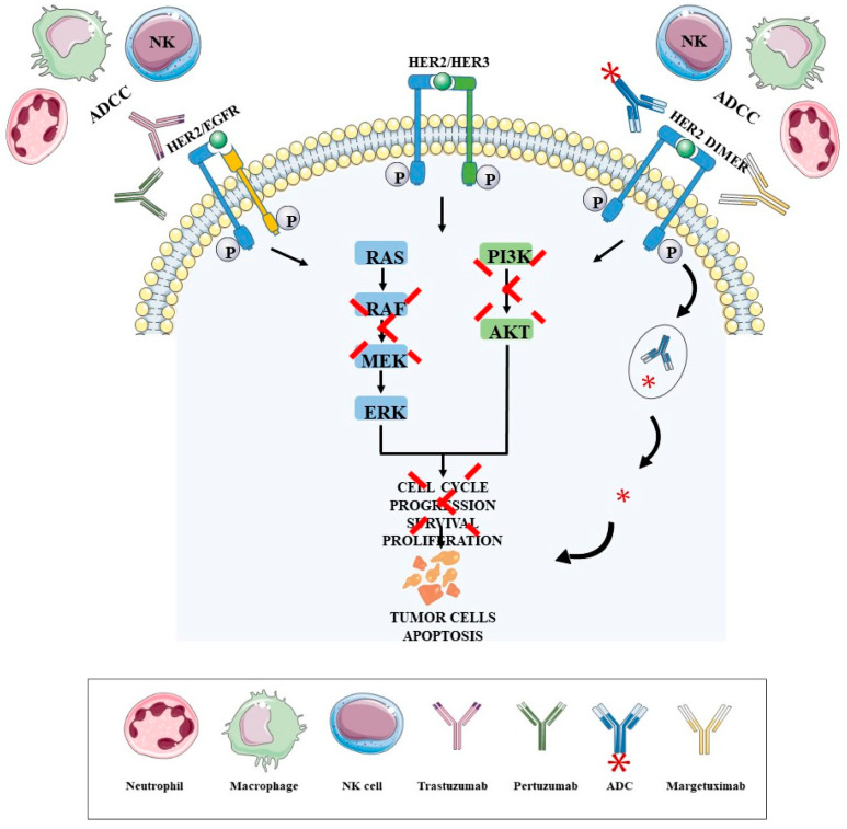
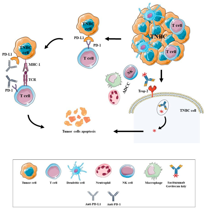
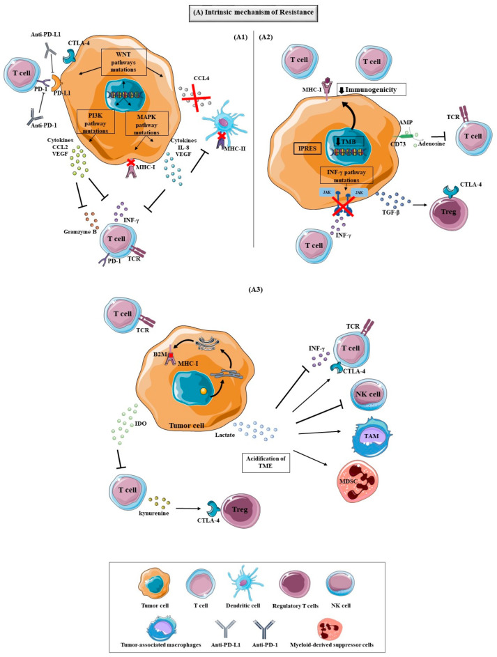
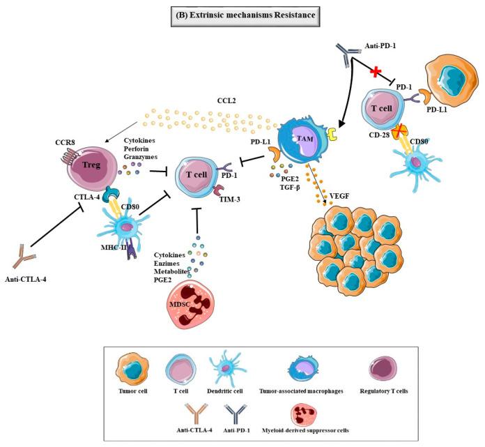
Breast Cancer (BC) is the second most frequent cause of cancer death among women worldwide and, although there have been significant advances in BC therapies, a significant percentage of patients develop metastasis and disease recurrence. Since BC was demonstrated to be an immunogenic tumor, immunotherapy has broken through as a significant therapy strategy against BC. Over the years, immunotherapy has improved the survival rate of HER2+ BC patients due to the approval of some monoclonal antibodies (mAbs) such as Trastuzumab, Pertuzumab and, recently, Margetuximab, along with the antibody-drug conjugates (ADC) Trastuzumab-Emtansine (T-DM1) and Trastuzumab Deruxtecan. Immune checkpoint inhibitors (ICI) showed promising efficacy in triple-negative breast cancer (TNBC) treatment, namely Atezolizumab and Pembrolizumab. Despite the success of immunotherapy, some patients do not respond to immunotherapy or those who respond to the treatment relapse or progress. The main causes of these adverse events are the complex, intrinsic or extrinsic resistance mechanisms. In this review, we address the different immunotherapy approaches approved for BC and some of the mechanisms responsible for resistance to immunotherapy.

Keywords: breast cancer; immunotherapy; therapeutic resistance.


Conflict of interest statement

The authors declare no conflict of interest.

Figures

Figure 1
Figure 1
Immunotherapy modalities approved as a treatment for patients with HER2 BC and their mechanisms of action. Trastuzumab, Pertuzumab and Margetuximab are the mAbs approved for HER2 BC therapy directed for HER2 protein in tumor cells, specifically for the domain II (Pertuzumab) and domain IV (Trastuzumab). When the mAbs attach, the HER2 protein blocks the dimerization of HER2 protein with the other receptors (HER2, HER3 or EGFR), inhibiting the activation of the HER2 protein and consequently the activation of the PI3K and MAPK pathways, which leads to an increase in cell cycle arrest, the suppression of cell growth and proliferation, causing the apoptosis of tumor cells. These mAbs can also activate both innate and adaptive immune systems, inducing the ADCC that allows to kill HER2-overexpressing cells via NK cells, macrophages and neutrophils, and eliciting an adaptive immune response based on HER2 presentation by MHC-I molecules to activate the anti-tumor activity of CTL. Other immunotherapies approved are the Trastuzumab-Emtansine and Trastuzumab Deruxtecan, that are an ADC who consists in a therapy that conjugates the Trastuzimab covalently conjugated with the cytotoxins DM1 (Trastuzumab-Emtansine), a microtubule polymerization inhibitor, and topoisomerase I inhibitor payload (Dxd) (Trastuzumab Deruxtecan), responsible for the inhibition of DNA replication. The Trastuzumab directs the ADC to the tumor cells that express HER2 protein and when the Trastuzumab binds to the HER2 protein the ADC goes into the cell by receptor-mediated endocytosis and the lysosomes degrade the Trastuzumab, realizing the DM1 or the Dxd. The DM1 inhibits the microtubule function and the Dxd inhibits the topoisomerase I, causing the cell cycle arrest and tumor cell apoptosis. Abbreviations are as follows: Antibody Dependent Cytotoxic Cell (ADCC), Antibody Drug Conjugate (ADC), Natural Killer (NK), Human Epidermal growth Receptor 2 (HER2), Human Epidermal Growth Receptor 3 (HER3), Human Epidermal Growth Factor Receptor (EGFR), Phosphatidylinositol-3-kinase (PI3K), Extracellular regulated kinases (ERK), Mitogen-activated protein kinase kinase (MEK), Protein kinase B (AKT).
Figure 2
Figure 2
Immunotherapy modalities approved as a treatment for patients with TNBC and their mechanism of action. The tumor cells can escape from host immune surveillance by the expression of immune checkpoint proteins, like PD-L1, that when bound to PD-1 expressed in active T cells, cause the decrease of T cell proliferation and survival. One of the strategies to restore antitumor immune responses and promote immune-mediated elimination of cancer cells are the ICI. Nowadays, Pembrolizumab (anti-PD-1) and Atezolizumab (anti-PD-L1) are two ICI approved for TNBC patients that when bound to their target blocks the interaction of PD-1 and PD-L1 and allows the CTL to eliminate the tumors cells. The Sacituzumab Govitecan-hziy was also approved as a treatment for TNBC patients, an ADC formed by the mAb hRS7 directed for antitrophoblast cell-surface antigen 2 (Trop-2), that was expressed in 90% of TNBC tumors, conjugated with SN-38, a topoisomerase I inhibitor. The hRS7 binds to the Trop-2 expressed in TNBC cells and the ADC goes into the cell by receptor-mediated endocytosis. The lysosomes degrade the mAb, realizing the SN-38 and inhibiting the topoisomerase I, causing the cell cycle arrest and tumor cell apoptosis. It was also demonstrated that Sacituzumab Govitecan-hziy promotes ADCC that kills the tumor cells that express the Trop-2. Abbreviations are as follows: Triple-Negative Breast Cancer (TNBC), Programmed Cell Death Protein-1 (PD-1), Programmed Cell Death Protein Ligand 1 (PD-L1), Major Histocompability Complex 1 (MHC-1), T cell Receptor (TCR), Antitrophoblast Cell-Surface Antigen 2 (Trop-2), Antibody Dependent Cytotoxic Cell (ADCC), Natural Killer (NK), Antibody Programmed Cell Death Protein-1 (Anti-PD-1), Antibody Programmed Cell Death Protein Ligand 1 (Anti-PD-L1).
Figure 3
Figure 3
Intrinsic mechanisms of tumor resistance to immunotherapy: (A1) The tumor cells have the ability to not respond to immunotherapy by the development of different mechanisms that have been described in different studies. One of the mechanisms that can lead to the resistance are the alterations of some signaling pathways in tumor cells that can cause the reduction or loss of antigens expression, preventing immune cell infiltration or function. The WNT signaling is one of these pathways that can be aberrantly activated in a variety of tumors and act in the tumor cell genesis, proliferation, invasiveness, metastatic and immune microenvironment regulation. In some studies, it was demonstrated that the WNT signaling can control the expression of PD-L1 and CTLA-4 and can reduce the expression of chemokine that attracts CD103+ DC (CCL4), decreasing the DC migration into the TME and contributing to the inhibition of activation and cytotoxic effects of T cells; The PI3K pathway is one of the responsible pathways that contributes to the development and growth of tumors and alterations in their intermediaries, like tumor suppressor protein phosphatase and tensin homolog (PTEN) loss can cause the release of anti-inflammatory cytokines, such as C-C Motif Chemokine Ligand 2 (CCL2) and VEGF that reduce the infiltration of CTL cells in tumors, decrease IFN-γ and granzyme B expression; MAPK signaling is very involved in the tumors development, wherein alterations in this pathway can suppress the expression of MHC-I and MHC-II and the induction of the expression of VEGF and a variety of inhibitory cytokines, such as the interleukin (IL)-8 proteins that inhibit T cell function and recruitment. (A2) The mutational load that tumors present can influence the expression of neoantigens and the tumors with low levels of TMB are considered less immunogenic, since they present low expression of neoantigens and a low number of TILs in TME, decreasing the immune response against the tumor. In a study with melanoma patients the expression of a group of transcriptomic signatures, referred as innate anti-PD-1 resistance signature (IPRES), by the tumors can regulate different processes, like mesenchymal transformation, angiogenesis, extracellular matrix remodeling and others, leading the tumor to not respond to anti-PD-1 therapy. The IFN- γ pathway is essential for the immune response, since the IFN- γ secreted by the T cells and APC directed for tumor cells contributes to the immune cell activation and regulation of T cell responses, and can also directly induce tumor cell death. The downregulation or mutations in the molecules involved in the IFN-γ signaling pathway, like JAK1/2 mutations by tumor cells, cause the survival of the tumor cells by resisting the antiproliferative effects of IFN-γ can lead to the tumor’s resistance to the antiproliferative effects of IFN-γ to escape the influence of IFN-γ; Tumor cells can realize immune-suppressive cytokines, like the transforming growth factor (TGF)-β, that can stimulate Treg cells, promoting the angiogenesis and immunosuppression of immune response. The CD73 can form adenosine by the dephosphorylation of adenosine monophosphate (AMP) and the adenosine and CD73 can inhibit the proliferation and function of T cells; (A3) the B2M protein is essential to transport MHC class I to the cell surface and mutation in this protein, disrupt the antigen presentation and, consequently, the recognition of CTL fails, which can lead to acquired resistance to immunotherapy. The IDO enzyme liberated by the tumor can reduce the T cell proliferation, inhibiting the activity of T-cell against tumor cells by the degradation of tryptophan into the kynurenine, and kynurenine stimulates the proliferation of Treg cells. The lactate produced by the tumor cells due to metabolic reprogramming can cause the acidification of the TME, affecting the IFN-γ production, NK activation and the amount of MDSC, causing the decrease of the immune response and contributing to the tumor growth. Abbreviations are as follows: T cell Receptor (TCR), Major Histocompability Complex 1 (MHC-I), Major Histocompability Complex 2 (MHC-II), Beta-2-Microglobulin (B2M), Interferon Gamma (IFN-γ), Cytotoxic T-Lymphocyte Associated Protein-4 (CTLA-4), Tumor Microenvironment (TME), Tumor-Associated Macrophages (TAM), Myeloid-Derived Suppressor Cells (MDSC), Regulatory T cells (Treg), Programmed Cell Death Protein-1 (PD-1), Programmed Cell Death Protein Ligand 1 (PD-L1), Antibody Programmed Cell Death Protein-1 (Anti-PD-1), Antibody Programmed Cell Death Protein Ligand 1 (Anti-PD-L1), Phosphatidylinositol-3-kinase (PI3K), Mitogen-Activated Protein Kinase (MAPK), C-C Motif Chemokine Ligand 2 (CCL2), Vascular Endothelial Growth Factor (VEGF), IInterleukin 8 (IL-8), Tumor Mutational Burden (TMB), Innate Anti-PD-1 Resistance Signature (IPRES), Adenosine Monophosphate (AMP), Transforming Growth Factor Gamma (TGF-β), Janus Kinase (JAK).
Figure 4
Figure 4
Extrinsic mechanisms of tumor resistance to immunotherapy. The presence of determinate components in TME, like immunosuppressive cells and some molecules, influenced by the tumor can lead to immunotherapy resistance. The Treg cells, characterized by the expression of the FoxP3, are one of these components and they can inhibit MHC molecules and CD80/CD86 on the surface of APC and secrete cytokines, perforin and granzymes that inhibit the activation and proliferation of T cells effectors and APC. The MDSC are a group of cells of type myeloid origin involved in TME, that have a potent immune-suppressive activity by inhibiting the T-cell function with the production of immunosuppressive metabolites, immunosuppressive cytokines, immunoactive enzymes, and immunosuppressive prostaglandin E2 (PGE2), contributing to the progression, invasion and metastasis of the tumor. The TAM (M1 and M2 macrophages) are other types of cells that can infiltrate around tumor cells, due to the action of VEGF and chemokines, and the overexpression of PD-L1, PGE2, TGF-β and CCL2 (promote the accumulation of Treg) affect responses to immunotherapy. In a study, it was demonstrated that TAM can capture anti–PD-1 from the surface of T cells, leading to the resistance of the PD-1 inhibitor. The expression of immune checkpoints can mediate tumor immune resistance, Tim-3 being one of these and expressed in T cells, whereby the upregulation of this immune checkpoint after the PD-1 blockade lead to the inhibition of the activation of T cells, decreasing immunotherapeutic response. The loss of expression of CD28, the homologue of CTLA-4 that promotes the activation of T cells, can cause the T cells lost capacity to proliferate and perform the normal activity, contributing to the immunotherapeutic resistance. Abbreviations are as follows: Cytotoxic T-Lymphocyte Associated Protein-4 (CTLA-4), Antibody Cytotoxic T-Lymphocyte Associated Protein-4 (Anti-CTLA-4), Tumor-Associated Macrophages (TAM), Myeloid-Derived Suppressor Cells (MDSC), Regulatory T cells (Treg), Programmed Cell Death Protein-1 (PD-1), Programmed Cell Death Protein Ligand 1 (PD-L1), Antibody Programmed Cell Death Protein-1 (Anti-PD-1), C-C Motif Chemokine Ligand 2 (CCL2), Vascular Endothelial Growth Factor (VEGF), Transforming Growth Factor Gamma (TGF-β), T cell Immunoglobulin 3 (TIM-3), Prostaglandin E2 (PGE2).
Figure 5
Figure 5
Known and hypothetical target pathways in breast cancer immunotherapeutic treatment. (A1) The immune checkpoint PD-1 is one of the mechanisms that can lead to immunotherapy resistance by the downstream of PI3K/AKT and MAPK signaling inhibition, causing the decrease of T cell proliferation and survival, cytokine production, as well as other effector functions. So, when the anti-PD-1 binds to PD-1 expressed in T cells stops the downstream of PI3K/AKT and MAPK signallings inhibition, promoting the activation and proliferation of T cells. Other two mechanisms that promote the activation and proliferation of T cells are the expression of CD28, the homologue of CTLA-4, and the TCR, when they bind to the CD80/86 in APC and MHC-I in tumor cells, respectively, activating the downstream of PI3K/AKT and MAPK signallings; (A2) The MAPK and PI3K pathways are the main signaling involved in BC, leading to the proliferation and progression of tumors and antibodies directed for the HER2 protein in tumor cells, inhibiting the activation of the HER2 protein and consequently the activation of the PI3K and MAPK pathways, leading to the apoptosis of tumor cells. Also, the tumor cells can express the immune checkpoint PD-L1 that acts as a mechanism of immunologic escape by the tumor cells when they bind to the PD-1 expressed in T cells. The PD-L1 expression in tumor cells can be stimulated by the INF-γ, WNT and PI3K pathways that can block PD-L1 by the antibodies directed for it, inhibiting the binding with PD-1 in T cells that allows the activation, proliferation and function of T cells against tumor cells; (B1) The expression of CTLA-4 by the T cells can limit the T cells activation through the competition with the CD28 for the ligands CD80/86 expressed in APC, and this immune checkpoint can be expressed as a mechanism to escape from host immune surveillance, decreasing the T cells functions. When the anti-CTLA-4 binds to CTLA-4, it allows the APC to bind to the CD28, promoting the activation and proliferation of T cells and immune response. Nowadays, no anti-CTLA-4 is approved as a treatment for BC patients, but some of them are in evaluation for BC patients; (B2) Other several mechanisms have been described as a resistance to immunotherapy, like the alterations of some signaling pathways. The alterations in the WNT signalling can lead to the expression of PD-L1 in tumor cells and reduce the expression of chemokine that attracts CD103+ DC (CCL4) by the high levels of β-catenin, decreasing the DC migration into the TME and contributing to the decrease of activation and cytotoxic effects of T cells. The IFN-γ is essential for the immune response when they are secreted by the T cells and APC binds to the receptor in tumor cells, contributing for the immune cell activation and regulation of T cell responses, and can also directly induce tumor cell death. The downregulation or mutations in the molecules involved in the IFN-γ signaling can cause the survival of the tumor cells by resisting the antiproliferative effects of IFN-γ. Tumor cells can also secrete some inhibitory molecules, like IDO enzyme that reduces the T cell proliferation and stimulates the proliferation of Treg cells, contributing to the immunotherapy resistance. The Treg cells also can express the CTLA-4, increasing the immune resistance, and the anti-CTLA-4 can reverse this affect by the mechanisms previously described. Abbreviations are as follows: Cytotoxic T-Lymphocyte Associated Protein-4 (CTLA-4), Antibody Cytotoxic T-Lymphocyte Associated Protein-4 (Anti-CTLA-4), Regulatory T cells (Treg), Programmed Cell Death Protein-1 (PD-1), Programmed Cell Death Protein Ligand 1 (PD-L1), Antibody Programmed Cell Death Protein-1 (Anti-PD-1), Human Epidermal growth Receptor 2 (HER2), Phosphatidylinositol-3-kinase (PI3K), Extracellular regulated kinases (ERK), Mitogen-activated protein kinase (MEK), Protein kinase B (AKT), signal transducers and activators of transcription (STAT), Janus Kinase (JAK), Major Histocompability Complex 1 (MHC-1), T cell Receptor (TCR), Antibody Programmed Cell Death Protein Ligand 1 (Anti-PD-L1), C-C Motif Chemokine Ligand 4 (CCL4), indoleamine-2,3-dioxygenase (IDO).

References

    1. Sivaganesh V., Promi N., Maher S., Peethambaran B. Emerging Immunotherapies against Novel Molecular Targets in Breast Cancer. Int. J. Mol. Sci. 2021;22:2433. doi: 10.3390/ijms22052433. - DOI - PMC - PubMed
    1. Tong C.W.S., Wu M., Cho W.C.S., To K.K.W. Recent Advances in the Treatment of Breast Cancer. Front. Oncol. 2018;8:227. doi: 10.3389/fonc.2018.00227. - DOI - PMC - PubMed
    1. Ellis H., Ma C.X. PI3K Inhibitors in Breast Cancer Therapy. Curr. Oncol. Rep. 2019;21:110. doi: 10.1007/s11912-019-0846-7. - DOI - PubMed
    1. Feng Y., Spezia M., Huang S., Yuan C., Zeng Z., Zhang L., Ji X., Liu W., Huang B., Luo W., et al. Breast cancer development and progression: Risk factors, cancer stem cells, signaling pathways, genomics, and molecular pathogenesis. Genes Dis. 2018;5:77–106. doi: 10.1016/j.gendis.2018.05.001. - DOI - PMC - PubMed
    1. Althoubaity F.K. Molecular classification of breast cancer: A retrospective cohort study. Ann. Med. Surg. 2019;49:44–48. doi: 10.1016/j.amsu.2019.11.021. - DOI - PMC - PubMed