. 2021 Nov 18;9(11):1715.
doi: 10.3390/biomedicines9111715.

Oxidized LDL Increase the Proinflammatory Profile of Human Visceral Adipocytes Produced by Hypoxia

Affiliations

Oxidized LDL Increase the Proinflammatory Profile of Human Visceral Adipocytes Produced by Hypoxia

Concepción Santiago-Fernández et al. Biomedicines. .

Abstract

Background: Little is known about the effects of hypoxia on scavenger receptors (SRs) levels in adipocytes. We analyzed the effect of morbid obesity and hypoxia on SRs and inflammation markers in human visceral adipocytes and whether ox-LDL modify the inflammatory profile produced by hypoxia.

Methods: We studied in 17 non-obese and 20 subjects with morbid obesity (MO) the mRNA expression of HIF-1α, SRs (LOX-1, MSR1, CL-P1 and CXCL16), IL6 and TNFα in visceral adipocytes and the effect of hypoxia with or without ox-LDL on visceral in vitro-differentiated adipocytes (VDA).

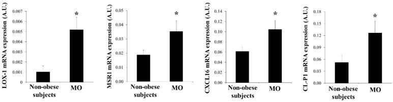
Results: HIF-1α, TNFα, IL6, LOX-1, MSR1 and CXCL16 expression in adipocytes was increased in MO when compared with those in non-obese subjects (p < 0.05). The expression of most of the inflammatory markers and SRs gene correlated with HIF-1α. In VDA, hypoxia increased TNFα, IL6, MSR1, CXCL16 and CL-P1 (p < 0.05) in non-obese subjects, and TNFα, IL6, MSR1 and CXCL16 (p < 0.05) in MO. Silencing HIF-1α prevented the increase of TNFα, IL6, LOX-1, MSR1, CL-P1 and CXCL16 expression (p < 0.05). The combination of hypoxia and ox-LDL produced higher TNFα expression (p = 0.041).

Conclusions: Morbid obesity and hypoxia increased SRs and inflammatory markers in visceral adipocytes. In a hypoxic state, ox-LDL increased the proinflammatory response of visceral adipocytes to hypoxia.

Keywords: adipocytes; hypoxia; morbid obesity; oxidized LDL; scavenger receptors.


Conflict of interest statement

The authors declare that they have no conflicts of interests.

Figures

Figure 1
Figure 1
(A) Levels of mRNA expression of HIF-1α, TNFα and IL6 in human visceral adipocytes from non-obese subjects and patients with morbid obesity (MO). Data are the mean ± SEM. * p < 0.05: significant differences between non-obese subjects and MO. (B) Significant correlation found in visceral mature adipocytes between the mRNA expression of HIF-1α and IL6.
Figure 2
Figure 2
Levels of mRNA expression of scavenger receptors in human visceral adipocytes from non-obese subjects and patients with morbid obesity (MO). Data are the mean ± SEM. * p < 0.05: significant differences between non-obese subjects and MO.
Figure 3
Figure 3
Significant correlations found in visceral mature adipocytes between the mRNA expression of scavenger receptors with HIF-1α and IL6.
Figure 4
Figure 4
Significant correlations found in visceral mature adipocytes between the mRNA expression of HIF-1α and scavenger receptors.
Figure 5
Figure 5
Levels of HIF-1α, GLUT1, IL6, TNFα and scavenger receptors mRNA expression of in vitro-differentiated adipocytes obtained from human mesenchymal stem cells of visceral adipose tissue (VAT) from non-obese subjects (n = 5) and patients with morbid obesity (n = 5) (MO) incubated in normoxia (■) and hypoxia (□) conditions for 24 h. Each treatment was performed in triplicate. Data are the mean ± SEM. * p < 0.05: significant differences between the normoxia and hypoxia conditions.
Figure 6
Figure 6
Levels of HIF-1α, GLUT1, TNFα, IL6 and scavenger receptors mRNA expression (%) of in vitro-differentiated adipocytes obtained from human mesenchymal stem cells of visceral adipose tissue (VAT) from non-obese subjects (n = 5) incubated in normoxia and hypoxia conditions when HIF-1α was silenced as described in the Materials and Methods section. Briefly, at day 15 of adipocyte differentiation, the transfection was made with a siRNA (s6539) and DharmaFECTTM Transfection Reagent. After 24 h in the normoxia condition, the cells were incubated for another 24 h in normoxia or hypoxia conditions. Cells were harvested and frozen at −80 °C until analysis. Each treatment was performed in triplicate. Data are the mean ± SEM. Results are shown as a percentage with regard to normoxia condition (100%) without hypoxia and HIF-1α silencing. * p < 0.05: significant differences between silenced and not silenced HIF-1α conditions. 1 p < 0.05: significant differences between normoxia and hypoxia conditions when HIF-1α is or is not silenced.
Figure 7
Figure 7
Levels of mRNA expression (%) of TNFα and IL6 of in vitro-differentiated adipocytes obtained from human mesenchymal stem cells of VAT from non-obese subjects (n = 5) incubated in hypoxia conditions with ox-LDL. Briefly, at day 15 of adipocyte differentiation, the cells were incubated for 48 h in normoxia or hypoxia conditions. After the first 24 h of incubation, 0 and 50 µg/mL of human ox-LDL were added. After another 24 h of incubation, the adipocytes were harvested and frozen at −80 °C until analysis. Each treatment was performed in triplicate. Data are the mean ± SEM. . Results are shown as a percentage with regard to normoxia condition (100%) without hypoxia and ox-LDL. a p < 0.05: significant differences respect to the normoxia condition without 50 µg/mL ox-LDL. 1 p < 0.05: significant differences between hypoxia and hypoxia + ox-LDL conditions.

References

    1. García-Fuentes E., Santiago-Fernández C., Gutiérrez-Repiso C., Mayas M.D., Oliva-Olivera W., Coín-Aragüez L., Alcaide J., Ocaña-Wilhelmi L., Vendrell J., Tinahones F.J., et al. Hypoxia is associated with a lower expression of genes involved in lipogenesis in visceral adipose tissue. J. Transl. Med. 2015;13:373. doi: 10.1186/s12967-015-0732-5. - DOI - PMC - PubMed
    1. Fernández-Sánchez A., Madrigal-Santillán E., Bautista M., Esquivel-Soto J., Morales-González A., Esquivel-Chirino C., Durante-Montiel I., Sánchez-Rivera G., Valadez-Vega C., Morales-González J.A. Inflammation, oxidative stress, and obesity. Int. J. Mol. Sci. 2011;12:3117–3132. doi: 10.3390/ijms12053117. - DOI - PMC - PubMed
    1. Yuan F., Guo Z.Z., Ji W.J., Ma Y.Q., Zhang Z., Zhou X., Li Y.M. BOLD-MRI evaluation of subcutaneous and visceral adipose tissue oxygenation status: Effect of dietary salt intake. Am. J. Transl. Res. 2015;7:598–606. - PMC - PubMed
    1. Trayhurn P., Wang B., Wood I.S. Hypoxia in adipose tissue: A basis for the dysregulation of tissue function in obesity? Br. J. Nutr. 2008;100:227–235. doi: 10.1017/S0007114508971282. - DOI - PubMed
    1. Trayhurn P., Wang B., Wood I.S. Hypoxia and the endocrine and signalling role of white adipose tissue. Arch. Physiol. Biochem. 2008;114:267–276. doi: 10.1080/13813450802306602. - DOI - PubMed