. 2021 Nov 9;22(22):12134.
doi: 10.3390/ijms222212134.

Xanthohumol Is a Potent Pan-Inhibitor of Coronaviruses Targeting Main Protease

Affiliations

Xanthohumol Is a Potent Pan-Inhibitor of Coronaviruses Targeting Main Protease

Yuxi Lin et al. Int J Mol Sci. .

Abstract

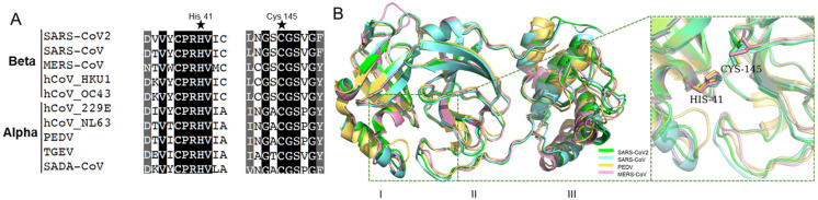
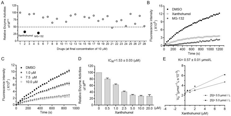
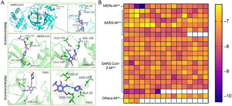
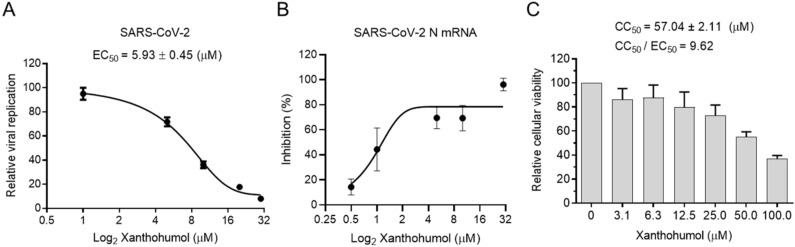
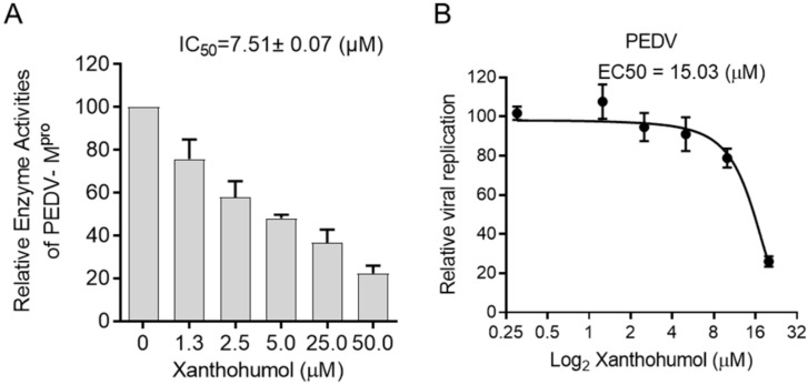
Coronaviruses cause diseases in humans and livestock. The SARS-CoV-2 is infecting millions of human beings, with high morbidity and mortality worldwide. The main protease (Mpro) of coronavirus plays a pivotal role in viral replication and transcription, which, in theory, is an attractive drug target for antiviral drug development. It has been extensively discussed whether Xanthohumol is able to help COVID-19 patients. Here, we report that Xanthohumol, a small molecule in clinical trials from hops (Humulus lupulus), was a potent pan-inhibitor for various coronaviruses by targeting Mpro, for example, betacoronavirus SARS-CoV-2 (IC50 value of 1.53 μM), and alphacoronavirus PEDV (IC50 value of 7.51 μM). Xanthohumol inhibited Mpro activities in the enzymatical assays, while pretreatment with Xanthohumol restricted the SARS-CoV-2 and PEDV replication in Vero-E6 cells. Therefore, Xanthohumol is a potent pan-inhibitor of coronaviruses and an excellent lead compound for further drug development.

Keywords: PEDV; SARS-CoV-2; Xanthohumol; coronavirus; natural product.


Conflict of interest statement

The authors declare no conflict of interest.

Figures

Figure 1
Figure 1
The conserved amino acid sequences and 3D structures of the Mpro catalytic domains for different coronaviruses. (A) Alignment of neighbor residues on both flanks of the pivotal residue His41, Cys145. (B) The three-dimensional (3D) structures of Mpro are highly conserved.
Figure 2
Figure 2
Xanthohumol inhibited hydrolase activities of SARS-CoV-2 Mpro in a dose-dependent manner. (A) Relative enzyme activities of SARS-CoV-2 Mpro in the presence of 10 μM compounds. (B) The kinetic curves in the presence of Xanthohumol, MG132, and the solvent, DMSO. Xanthohumol and MG132 were added to the final concentration of 20 μM. (C) Xanthohumol inhibited Mpro dose-dependently. (D) The IC50 value of Xanthohumol. (E) The Ki value of Xanthohumol on SARS-CoV-2 Mpro. Data are shown as means ± standard error of mean (SEM) from three independent experiments.
Figure 3
Figure 3
Xanthohumol is a potent pan-inhibitor of coronaviral Mpro. (A) The interaction pattern between Xanthohumol and indicated Mpro. The Mpro was shown in cyan and green. The Xanthohumol was presented in purple. (B) The affinity diagrams between Xanthohumol and different Mpro. Mpro structures were downloaded from PDB, and the PDB ID and detailed docking scores are shown in Supplementary Data.
Figure 4
Figure 4
Xanthohumol inhibited SARS-CoV-2 replication in Vero-E6 cells. (A) Xanthohumol-inhibited SARS-CoV-2. Vero-E6 cells were pretreated with compounds at indicated concentrations for 1 h, and then infected with recombinant SARS-CoV-2 mNeonGreen virus (MOI = 0.5) for 24 h. The green fluorescence was scanned as described. The EC50 values of Xanthohumol were calculated and are shown. (B) Xanthohumol reduced viral RNA loads. SARS-CoV-2 N mRNA was measured with qRT-PCR and normalized to GAPDH. (C) Cytotoxicities of Xanthohumol. Data are shown as means ± SEM from three independent experiments.
Figure 5
Figure 5
Xanthohumol inhibited PEDV Mpro and PEDV in vitro. (A) Xanthohumol inhibited PEDV Mpro. (B) Xanthohumol inhibited PEDV replication in vitro. Vero-E6 cells were pretreated with the indicated concentration of Xanthohumol for 1 h and infected with PEDV (MOI = 1). Data are shown as means ± SEM from three independent experiments.

Similar articles

Cited by

References

    1. Cui J., Li F., Shi Z.L. Origin and evolution of pathogenic coronaviruses. Nat. Rev. Microbiol. 2019;17:181–192. doi: 10.1038/s41579-018-0118-9. - DOI - PMC - PubMed
    1. Kirtipal N., Bharadwaj S., Kang S.G. Evolution. From SARS to SARS-CoV-2, insights on structure, pathogenicity and immunity aspects of pandemic human coronaviruses. Infect. Genet. Evol. 2020;85:104502. doi: 10.1016/j.meegid.2020.104502. - DOI - PMC - PubMed
    1. Tizard I.R. Vaccination against coronaviruses in domestic animals. Vaccine. 2020;38:5123–5130. doi: 10.1016/j.vaccine.2020.06.026. - DOI - PMC - PubMed
    1. Edwards C.E., Yount B.L., Graham R.L., Leist S.R., Hou Y.J., Dinnon K.H., Sims A.C., Swanstrom J., Gully K., Scobey T.D., et al. Swine acute diarrhea syndrome coronavirus replication in primary human cells reveals potential susceptibility to infection. Proc. Natl. Acad. Sci. USA. 2020;117:26915–26925. doi: 10.1073/pnas.2001046117. - DOI - PMC - PubMed
    1. Lee P.I., Hsueh P.R. Emerging threats from zoonotic coronaviruses-from SARS and MERS to 2019-nCoV. J. Microbiol. Immunol. Infect. 2020;53:365–367. doi: 10.1016/j.jmii.2020.02.001. - DOI - PMC - PubMed

MeSH terms

LinkOut - more resources