Review
. 2021 Nov 10;22(22):12149.
doi: 10.3390/ijms222212149.

Immune Tolerance in the Oral Mucosa

Affiliations
Review

Immune Tolerance in the Oral Mucosa

Hector F Pelaez-Prestel et al. Int J Mol Sci. .

Abstract

The oral mucosa is a site of intense immune activity, where a large variety of immune cells meet to provide a first line of defense against pathogenic organisms. Interestingly, the oral mucosa is exposed to a plethora of antigens from food and commensal bacteria that must be tolerated. The mechanisms that enable this tolerance are not yet fully defined. Many works have focused on active immune mechanisms involving dendritic and regulatory T cells. However, epithelial cells also make a major contribution to tolerance by influencing both innate and adaptive immunity. Therefore, the tolerogenic mechanisms concurring in the oral mucosa are intertwined. Here, we review them systematically, paying special attention to the role of oral epithelial cells.

Keywords: T cell; dendritic cell; epithelial cell; mucosa; oral; tolerance.


Conflict of interest statement

The authors declare no conflict of interest.

Figures

Figure 1
Figure 1
The oral mucosa and associated lymphoid structures. The oral mucosa is lined by stratified squamous epithelia of varying thickness that can be topped by a keratin layer. Some areas related with mastication (a) are thick and highly keratinized, while other zones, such as the lining mucosa (b), are thinner and feature no keratin layer. Oral epithelial cells (OECs) are derived from local progenitor cells such as oral mucosal lamina propria-progenitor cells (OMLP-PCs). Resident mesenchymal stem cells (MSC) can also differentiate to epithelial cells as well as many other cell types. Resident dendritic cells (DC) can capture these antigens and migrate to secondary lymphoid tissues, including the tonsils (c) and proximal lymph nodes (d), where adaptive immune responses are elicited (inductive sites). CD103+ CCR4+ Tregs are induced in draining secondary lymphoid tissues (d) and migrate to the effector site, where they suppress immune responses when needed via CTLA-4 and LAG-3 expression and IL-10 and TGFβ production (b). The tonsils are exposed to a constant bombardment of antigens from innocuous sources, such as food, that need to be tolerated. Regulatory follicular T cells (TFR) as well as CD25+ TFH cells also control GC size and class switching by releasing IL-10 (c).
Figure 2
Figure 2
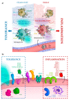
Immune modulation by OECs. (a) The fate of the DCs depends on the environment. Under homeostatic conditions, DCs exhibit a tolerogenic phenotype imprinted by surroundings OECs. Low expression of co-stimulatory molecules, high expression of inhibitory molecules and release of tolerogenic cytokines characterize tolerogonic DCs. Moreover, these cells can induce pTreg differentiation. However, threatening conditions, mainly the invasion of the mucosa by pathogenic species may change this environment, and freshly recruited DCs or pre-DCs (immature DCs) develop an inflammatory phenotype able to promote a non-tolerogenic immune response. (b) Molecular immunomodulatory arsenal of oral epithelial cells (OECs). OECs express different innate receptors, such as TLRs, NLRs, or inflammasome components that allow them to sense the environment and act in response to pathogens or damage. In addition, OECs modulate the immune response by secreting soluble factors, such as cytokines, prostaglandins, and kynurenines, and by expressing proteins at the cell surface that allow direct interaction with other immune cells, as indicated in Section 5.
Figure 3
Figure 3
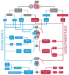
Mechanisms of immune tolerance in the oral mucosa. OECs secrete soluble molecules and express surface proteins that modulate immune responses. They condition DCs to become tolerogenic (blue), which promotes Treg differentiation, but can also promote the recruitment of inflammatory DCs (red). OECs also condition T cell responses by suppressing T cell responses (blue).

References

    1. Wertz P.W. Roles of Lipids in the Permeability Barriers of Skin and Oral Mucosa. Int. J. Mol. Sci. 2021;22:5229. doi: 10.3390/ijms22105229. - DOI - PMC - PubMed
    1. Gartner L.P. Oral anatomy and tissue types. Semin. Dermatol. 1994;13:68–73. - PubMed
    1. Nanci A. Ten Cate’s Oral Histology: Development, Structure, and Function. 8th ed. Elsevier; Montreal, QC, Canada: 2012.
    1. Mistretta C.M., Kumari A. Tongue and Taste Organ Biology and Function: Homeostasis Maintained by Hedgehog Signaling. Annu. Rev. Physiol. 2017;79:335–356. doi: 10.1146/annurev-physiol-022516-034202. - DOI - PMC - PubMed
    1. van der Bilt A., Engelen L., Pereira L.J., van der Glas H.W., Abbink J.H. Oral physiology and mastication. Physiol. Behav. 2006;89:22–27. doi: 10.1016/j.physbeh.2006.01.025. - DOI - PubMed

LinkOut - more resources