. 2021 Nov 10;22(22):12158.
doi: 10.3390/ijms222212158.

Tau Cleavage Contributes to Cognitive Dysfunction in Strepto-Zotocin-Induced Sporadic Alzheimer's Disease (sAD) Mouse Model

Affiliations

Tau Cleavage Contributes to Cognitive Dysfunction in Strepto-Zotocin-Induced Sporadic Alzheimer's Disease (sAD) Mouse Model

Valentina Latina et al. Int J Mol Sci. .

Abstract

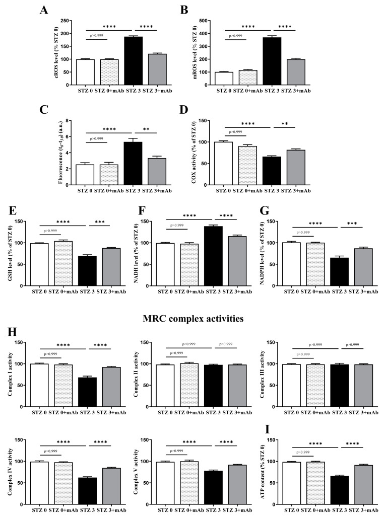
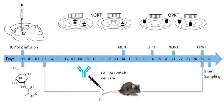
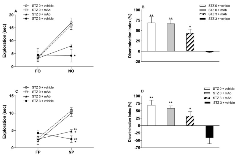
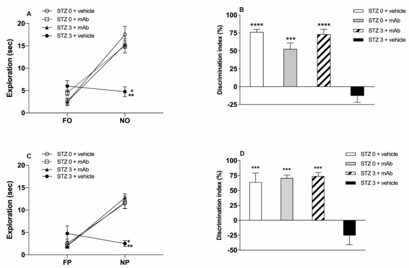
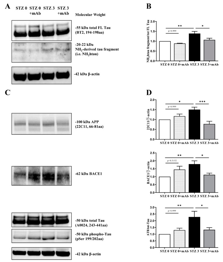
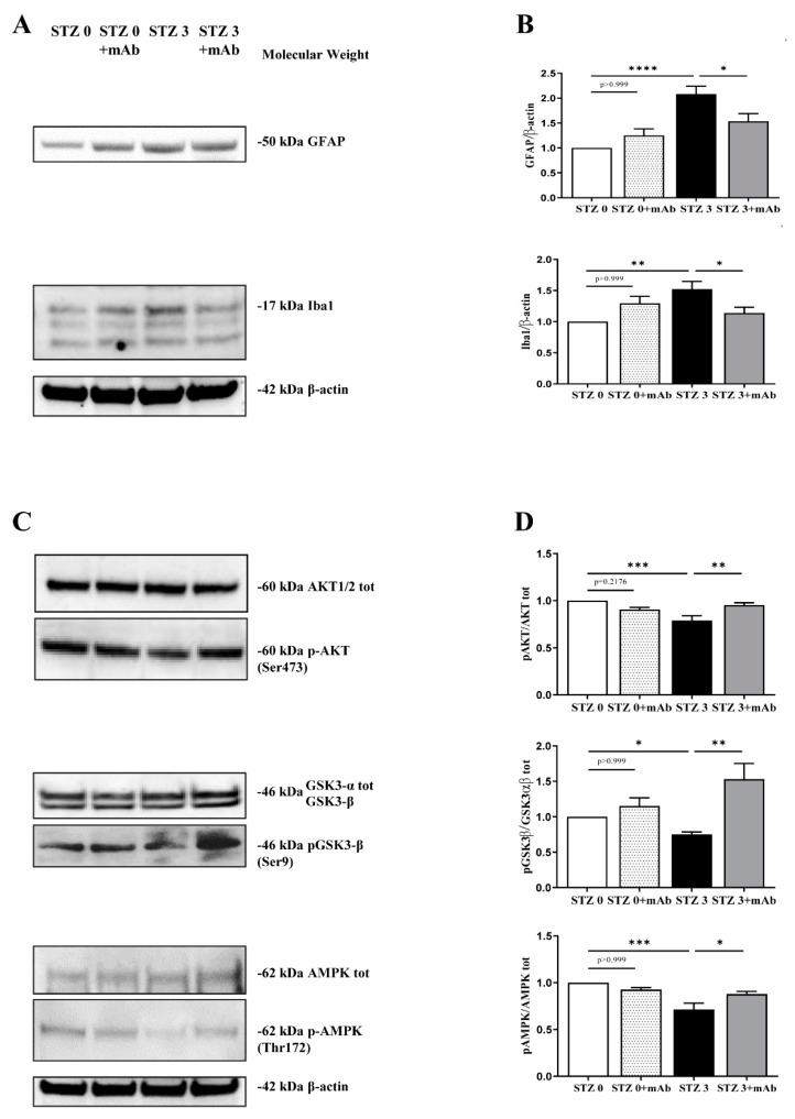
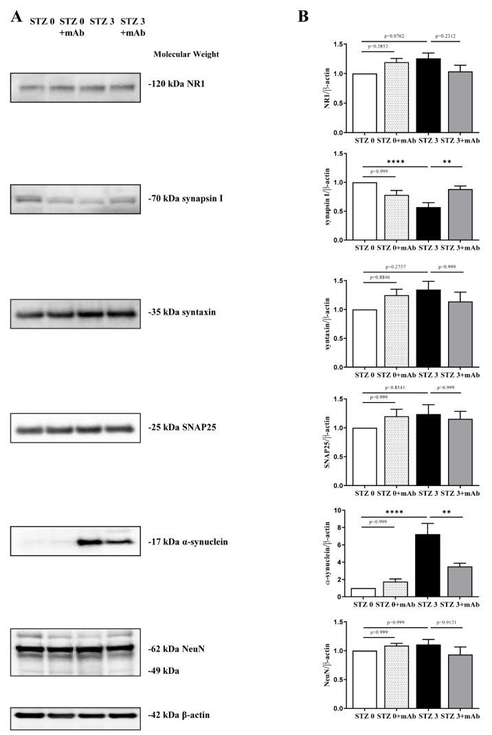
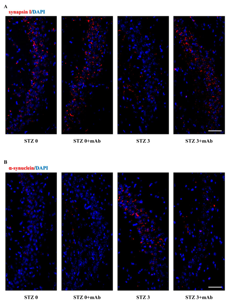
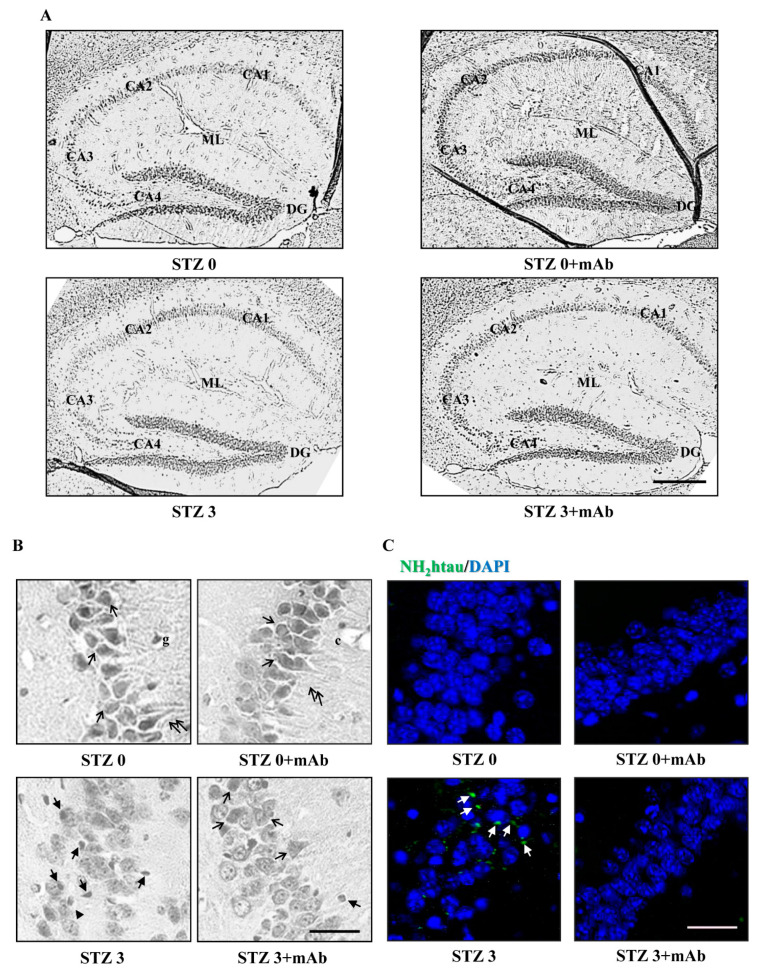
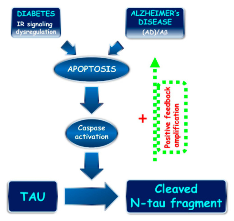
Tau cleavage plays a crucial role in the onset and progression of Alzheimer's Disease (AD), a widespread neurodegenerative disease whose incidence is expected to increase in the next years. While genetic and familial forms of AD (fAD) occurring early in life represent less than 1%, the sporadic and late-onset ones (sAD) are the most common, with ageing being an important risk factor. Intracerebroventricular (ICV) infusion of streptozotocin (STZ)-a compound used in the systemic induction of diabetes due to its ability to damage the pancreatic β cells and to induce insulin resistance-mimics in rodents several behavioral, molecular and histopathological hallmarks of sAD, including memory/learning disturbance, amyloid-β (Aβ) accumulation, tau hyperphosphorylation, oxidative stress and brain glucose hypometabolism. We have demonstrated that pathological truncation of tau at its N-terminal domain occurs into hippocampi from two well-established transgenic lines of fAD animal models, such as Tg2576 and 3xTg mice, and that it's in vivo neutralization via intravenous (i.v.) administration of the cleavage-specific anti-tau 12A12 monoclonal antibody (mAb) is strongly neuroprotective. Here, we report the therapeutic efficacy of 12A12mAb in STZ-infused mice after 14 days (short-term immunization, STIR) and 21 days (long-term immunization regimen, LTIR) of i.v. delivery. A virtually complete recovery was detected after three weeks of 12A12mAb immunization in both novel object recognition test (NORT) and object place recognition task (OPRT). Consistently, three weeks of this immunization regimen relieved in hippocampi from ICV-STZ mice the AD-like up-regulation of amyloid precursor protein (APP), the tau hyperphosphorylation and neuroinflammation, likely due to modulation of the PI3K/AKT/GSK3-β axis and the AMP-activated protein kinase (AMPK) activities. Cerebral oxidative stress, mitochondrial impairment, synaptic and histological alterations occurring in STZ-infused mice were also strongly attenuated by 12A12mAb delivery. These results further strengthen the causal role of N-terminal tau cleavage in AD pathogenesis and indicate that its specific neutralization by non-invasive administration of 12A12mAb can be a therapeutic option for both fAD and sAD patients, as well as for those showing type 2 diabetes as a comorbidity.

Keywords: cognitive/memory impairment; immunotherapy; neuroinflammation; non-transgenic Alzheimer’s Disease mouse model; oxidative stress; streptozotocin (STZ); tau cleavage.


Conflict of interest statement

Authors have no conflict of interest to be disclosed. All authors have seen and approved the manuscript.

Figures

Figure 6
Figure 6
Overt oxidative stress and mitochondrial deficits occurring in brains from ICV-STZ mice are recovered by 12A12mAb immunization. (AI) Cortical homogenates from animals of four experimental groups (STZ 0 plus vehicle; STZ 0 plus mAb; STZ 3 plus vehicle; STZ 3 plus mAb) were assessed for oxidative stress and oxidative phosphorylation. In (A,B): ROS level. Cytosolic and mitochondrial O2−• productions were detected according to the adrenochrome method (A) and by using the MitoSOX dye (B), respectively. The O2−• level value was expressed as percentage of STZ 0 plus vehicle, to which value 100 was given. In C–D: Lipid peroxidation and COX activity. Index of lipid peroxidation (C) are expressed as fluorescence values at the beginning of the reaction (t0) minus the value measured after 30 min (t30), accordingly to [66]. COX activity (D) was determined by measuring polarographically O2 − • consumption in the oxygen electrode chamber [74]. In (EG): GSH, NADH and NADPH level determinations were calculated as in [68]. In (H): MRC complex activities: the activities of Complex I (NADH:ubiquinone oxidoreductase), Complex II (succinate:ubiquinone oxidoreductase), Complex III (cytochrome c reductase), Complex IV (cytochrome c oxidase) and Complex V (ATP synthase) [39]. In (I): Cellular ATP content [39]. Values are from at least three independent experiments and statistically significant differences were calculated by one-way ANOVA followed by Bonferroni’s post-hoc test for multiple comparison among more than two groups. p < 0.05 was accepted as statistically significant (** p < 0.01; *** p < 0.0005; **** p < 0.0001).
Figure 1
Figure 1
Illustration of experimental design. Establishment of streptozotocin (STZ)-induced sporadic Alzheimer’s Disease (sAD) mouse model, immunization regimen and experimental procedures are shown. The mice were divided into four groups: STZ 0 plus vehicle; STZ 0 plus mAb; STZ 3 plus vehicle; STZ 3 plus mAb and housed in groups. The dose of 12A12mAb administered to mice in this study was calculated according to [1,39] and administered by intravenous injection into the lateral caudal vein on two alternated days up to 3 weeks.
Figure 2
Figure 2
Novel object recognition test (NORT) and object place recognition task (OPRT) cognitive scores of ICV-STZ mice after short-term immunization regimen with 12A12mAb. (A) The panel depicts the level of exploration (sec) during the NORT test phase. Unlike naïve ICV-STZ mice (STZ 3 plus vehicle), both groups of non-STZ-infused mice (STZ 0 plus vehicle and STZ 0 plus mAb) significantly directed their exploratory activity towards the novel object (NO), irrespective of mAb treatment (* p < 0.05). ICV-STZ mice that underwent i.v. delivery of 12A12mAb (STZ 3 plus mAb) showed a trend of increase of NO exploration which, however, was not statistically different from that of their non-immunized counterparts (STZ 3 plus vehicle vs. STZ 3 plus mAb, n.s.). (B) The panel depicts the level of DI during the NORT test phase, showing that naïve ICV-STZ mice (STZ 3 plus vehicle) were severely cognitively impaired, being unable to discriminate between the familial (FO) and NO objects (i.e., negative DI index). On the contrary, both groups of non-STZ mice (STZ 0 plus vehicle and STZ 0 plus mAb) showed significantly higher DI (** p < 0.01) in comparison with naïve ICV-STZ mice (STZ 3 plus vehicle). An intermediate level of DI was showed by ICV-STZ mice that underwent immunization (STZ 3 plus mAb vs. STZ 3 plus vehicle, * p < 0.05). (C) The panel depicts the level of exploration (sec) during the OPRT test phase. Naïve ICV-STZ mice (STZ 3 plus vehicle) did not explore the NP, while both groups of non-STZ-infused mice significantly directed their exploratory activity towards the NP, irrespective of mAb treatment (STZ 3 plus vehicle vs STZ 0 plus mAb, ** p < 0.01; STZ 3 plus vehicle vs. STZ 0 plus vehicle, * p < 0.05). ICV-STZ mice that underwent immunization (STZ 3 plus mAb) showed an increase of NP exploration which was, however, still lower than that of non-STZ mice (STZ 3 plus mAb vs. STZ 0 plus mAb, ** p < 0.01; STZ 3 plus mAb vs. STZ 0 plus vehicle, * p < 0.05). (D) The panel depicts the level of DI during the OPRT test phase, showing the complete lack of discrimination (i.e., negative DI index) exhibited by naïve ICV-STZ mice (STZ 3 plus vehicle). In contrast, both groups of non-STZ mice (STZ 0 plus vehicle and STZ 0 plus mAb) displayed significantly higher DI (** p < 0.01) in comparison with naïve ICV-STZ mice. ICV-STZ mice that underwent i.v. delivery of 12A12mAb showed an intermediate level of DI as compared to non-immunized counterpart (STZ 3 plus mAb vs. STZ 3 plus vehicle, * p < 0.05).
Figure 3
Figure 3
NORT and OPRT cognitive scores of ICV-STZ mice after long-term immunization regimen with 12A12mAb. (A) The panel depicts the level of exploration (sec) during the NORT test phase. Unlike naïve ICV-STZ mice (STZ 3 plus vehicle), both non-STZ-infused mice (STZ 0 plus vehicle and STZ 0 plus mAb) as well as ICV-STZ mice that underwent immunization with 12A12mAb (STZ 3 plus mAb) significantly oriented their exploratory activity towards the NO (STZ 3 plus vehicle vs. STZ 0 plus mAb and STZ 0 plus vehicle, ** p < 0.01; STZ 3 plus vehicle vs. STZ 3 plus mAb, * p < 0.05). (B) The panel depicts the level of DI during the NORT test phase, showing that non-immunized ICV-STZ mice (STZ 3 plus vehicle) were severely cognitively impaired, being unable to discriminate between the FO and NO objects (i.e., negative DI index). On the contrary, both STZ 0 plus vehicle and STZ 3 plus mAb groups, and also STZ 0 plus mAb mice showed significantly higher DI in comparison with non-immunized ICV-STZ mice (STZ 0 plus vehicle and STZ 3 plus mAb vs STZ 3 plus vehicle, **** p < 0.0001; STZ 0 plus mAb vs STZ 3 plus vehicle, *** p < 0.0005). (C) The panel depicts the level of exploration (sec) during the OPRT test phase. Naïve ICV-STZ mice (STZ 3 plus vehicle) did not explore the novel place (NP). On the contrary, both non-STZ mice (STZ 0 plus vehicle and STZ 0 plus mAb) and ICV-STZ-infused mice that underwent immunization (STZ 3 plus mAb) significantly oriented their exploratory activity towards the NP (STZ 3 plus vehicle vs. STZ 0 plus mAb and STZ 0 plus vehicle, * p < 0.05; STZ 3 plus vehicle vs. STZ 3 plus mAb, ** p < 0.01). (D) The panel depicts the level of DI during the OPRT test phase, showing the complete lack of discrimination (i.e., negative DI index) of naïve ICV-STZ mice (STZ 3 plus vehicle). On the contrary, both groups of non-STZ mice (STZ 0 plus vehicle and STZ 0 plus mAb) and ICV-STZ immunized mice (STZ 3 plus mAb) showed significantly higher DI (*** p < 0.0005) in comparison with naïve counterpart (STZ 3 plus vehicle).
Figure 4
Figure 4
The 12A12mAb-mediated neutralization of the N-terminal tau cleavage in brains from ICV-STZ mice is associated with reduction in AD-linked, pathologically relevant protein hallmarks. (AC) Hippocampal homogenates from animals of four experimental groups (STZ 0 plus vehicle; STZ 0 plus mAb; STZ 3 plus vehicle; STZ 3 plus mAb) were analyzed by Western blotting by developing with antibodies reported alongside the blot; (BD) semi-quantitative densitometry of the intensity signals of bands was carried out following normalization with β-actin level used as loading control. Arrows on the right side indicate the molecular weight (kDa) of bands calculated from migration of standard proteins. Values are from at least three independent experiments and statistically significant differences were calculated by one-way ANOVA followed by Bonferroni’s post-hoc test for multiple comparison among more than two groups. p < 0.05 was accepted as statistically significant (* p < 0.05; ** p < 0.01; *** p < 0.0005).
Figure 5
Figure 5
Effect of 12A12mAb immunization on cerebral neuroinflammation and dysregulation in several signal transduction pathways underlying the AD-like phenotype of ICV-STZ mice. (AC) Hippocampal homogenates from animals of four experimental groups (STZ 0 plus vehicle; STZ 0 plus mAb; STZ 3 plus vehicle; STZ 3 plus mAb) were analyzed by Western blotting by developing with antibodies reported alongside the blots. (BD) Semi-quantitative densitometry of the intensity signals of bands was carried out following normalization with β-actin level used as loading control. Arrows on the right side indicate the molecular weight (kDa) of bands calculated from migration of standard proteins. Values are from at least three independent experiments and statistically significant differences were calculated by one-way ANOVA followed by Bonferroni’s post-hoc test for multiple comparison among more than two groups. p < 0.05 was accepted as statistically significant (* p < 0.05; ** p < 0.01; *** p < 0.0005; **** p < 0.0001).
Figure 7
Figure 7
STZ-induced imbalanced expression of key synaptic proteins in hippocampus is normalized following 12A12mAb administration. (A) Hippocampal homogenates from animals of four experimental groups (STZ 0 plus vehicle; STZ 0 plus mAb; STZ 3 plus vehicle; STZ 3 plus mAb) were analyzed by Western blotting by developing with antibodies against several synaptic, including synapsin I, SNAP25 α-synuclein, syntaxin, N-Methyl-d-aspartate (NMDA) receptor subunit NR1, NeuN. (B) Semi-quantitative densitometry of the intensity signals of bands was carried out following normalization with β-actin level used as loading control. Arrows on the right side indicate the molecular weight (kDa) of bands calculated from migration of standard proteins. Values are from at least three independent experiments and statistically significant differences were calculated by one-way ANOVA followed by Bonferroni’s post-hoc test for multiple comparison among more than two groups. p < 0.05 was accepted as statistically significant; ** p < 0.01; **** p < 0.0001).
Figure 8
Figure 8
Effect of 12A12mAb immunization on synaptic changes of synapsin I and α-synuclein in the hippocampus of ICV-STZ mice. (A,B) Representative images (n = 3) of immunofluorescence analysis (20×) showing the punctate staining of the synapsin I (A) and α-synuclein (B) (red channel) in hippocampal CA2/CA3 regions from animals of four experimental groups (STZ 0 plus vehicle; STZ 0 plus mAb; STZ 3 plus vehicle; STZ 3 plus mAb). Nuclei were counterstained with DAPI (blue channel). Scale bar = 20 µm.
Figure 9
Figure 9
Histopathological alterations in the hippocampus of ICV-STZ mice are mitigated following 12A12mAb-mediated neutralization of the NH2htau. (A) Representative images of hippocampal slices showing hematoxylin and eosin staining under examination with a light electric microscope at 4×. Scale bar = 50 μm. (B) 40× magnification of CA3 subfield. Notice that STZ alters the laminar organization and significantly increases the number of degenerating neurons with apoptotic disintegration of the nucleus (black arrows with heads). Following 12A12mAb treatment, neuroprotection is evident as shown by the presence of well-preserved hippocampal cyto-architecture characterized by viable neurons with healthy, not-damaged nuclei (arrows). Scale bar = 10 μm. (C) Immunofluorescence analysis (n = 4) at 40× magnification showing the distribution/expression of the NH2htau peptide (green channel) in hippocampal CA3 regions from animals of four experimental groups (STZ 0 plus vehicle; STZ 0 plus mAb; STZ 3 plus vehicle; STZ 3 plus mAb). Nuclei were counterstained with DAPI (blue channel). Notice that, unlike not-immunized mice, the laminar organization/integrity is well preserved in STZ-treated hippocampi following 12A12mAb treatment in correlation with a significant diminution in signal of the NH2htau (white arrows). Scale bar = 10 µm.
Figure 10
Figure 10
N-terminal tau truncation: a common mechanism linking sporadic AD pathogenesis and diabetes. Proposed role of N-terminal tau cleavage correlating sAD and diabetes. An imbalance in IR signaling (diabetes) and APP/Aβ dysmetabolisms (AD) trigger the apoptotic pathway, including caspase(s) activation [42,112] and consequent N-terminal tau truncation with generation of the 20–22 kDa NH2htau peptide. The NH2htau promotes the APP/Aβ misprocessing [115,116] and perpetuates/amplifies the neurodegenerative process by means of feedback forward mechanism.

References

    1. Corsetti V., Borreca A., Latina V., Giacovazzo G., Pignataro A., Krashia P., Natale F., Cocco S., Rinaudo M., Malerba F., et al. Passive immunotherapy for N-truncated tau ameliorates the cognitive deficits in two mouse Alzheimer’s disease models. Brain Commun. 2020;2:fcaa039. doi: 10.1093/braincomms/fcaa039. - DOI - PMC - PubMed
    1. Hoyer S. Glucose metabolism and insulin receptor signal transduction in Alzheimer disease. Eur. J. Pharmacol. 2004;490:115–125. doi: 10.1016/j.ejphar.2004.02.049. - DOI - PubMed
    1. Mosconi L. Brain glucose metabolism in the early and specific diagnosis of Alzheimer’s disease. FDG-PET studies in MCI and AD. Eur. J. Nucl. Med. Mol. Imaging. 2005;32:486–510. doi: 10.1007/s00259-005-1762-7. - DOI - PubMed
    1. Flores-Cuadra J.A., Madrid A., Fernández P.L., Pérez-Lao A.R., Oviedo D.C., Britton G.B., Carreira M.B. Critical Review of the Alzheimer’s Disease Non-Transgenic Models: Can They Contribute to Disease Treatment? J. Alzheimer’s Dis. 2021;82:S227–S250. doi: 10.3233/JAD-200870. - DOI - PubMed
    1. Steen E., Terry B.M., Rivera E.J., Cannon J.L., Neely T.R., Tavares R., Xu X.J., Wands J.R., de la Monte S.M. Impaired insulin and insulin-like growth factor expression and signaling mechanisms in Alzheimer’s disease—Is this type 3 diabetes? J. Alzheimer’s Dis. 2005;7:63–80. doi: 10.3233/JAD-2005-7107. - DOI - PubMed