. 2021 Nov 10;22(22):12184.
doi: 10.3390/ijms222212184.

Patient-Specific iPSC-Derived Neural Differentiated and Hepatocyte-like Cells, Carrying the Compound Heterozygous Mutation p.V1023Sfs*15/p.G992R, Present the "Variant" Biochemical Phenotype of Niemann-Pick Type C1 Disease

Affiliations

Patient-Specific iPSC-Derived Neural Differentiated and Hepatocyte-like Cells, Carrying the Compound Heterozygous Mutation p.V1023Sfs*15/p.G992R, Present the "Variant" Biochemical Phenotype of Niemann-Pick Type C1 Disease

Christin Völkner et al. Int J Mol Sci. .

Abstract

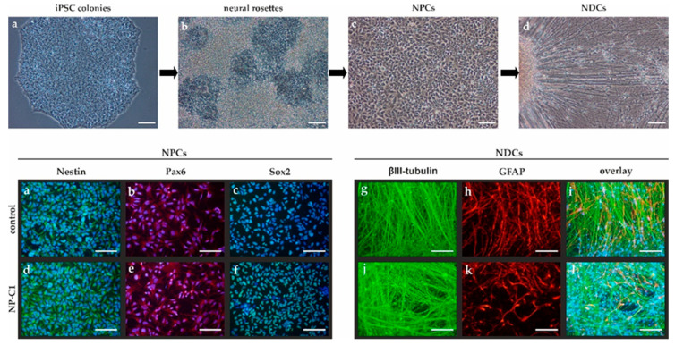
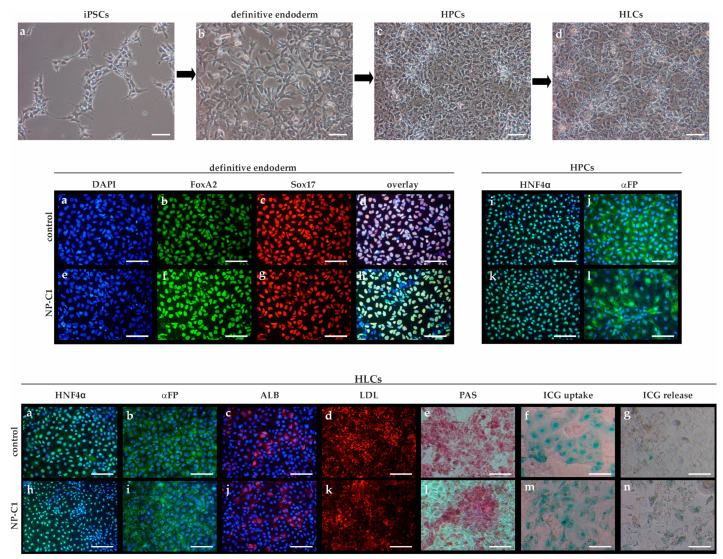
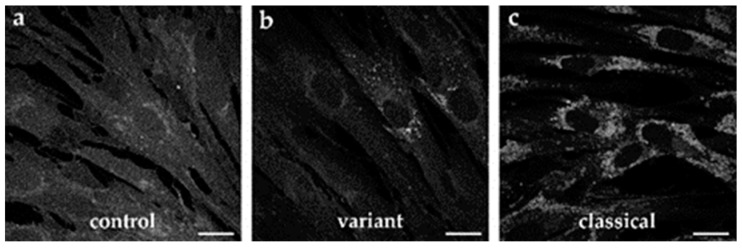
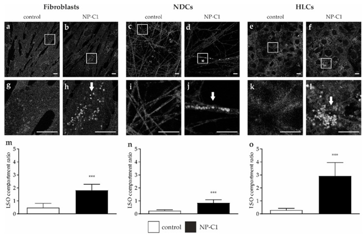
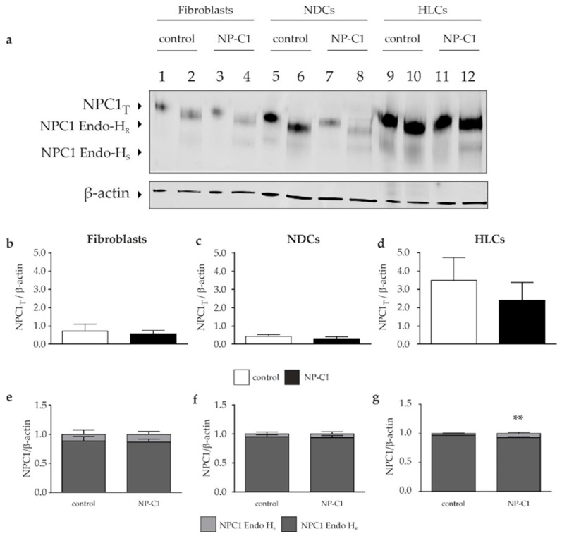
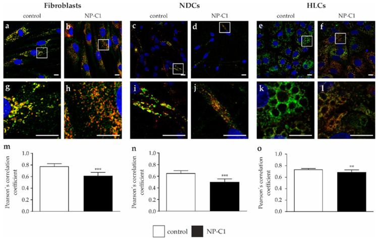
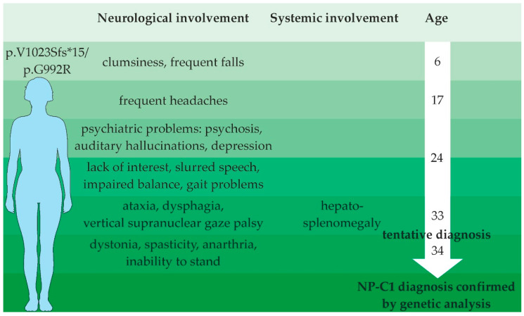
Niemann-Pick disease type C1 (NP-C1) is a rare lysosomal storage disorder caused by autosomal recessive mutations in the NPC1 gene. Patients display a wide spectrum on the clinical as well as on the molecular level, wherein a so-called "variant" biochemical phenotype can be observed. Here, we report an in vitro analysis of fibroblasts obtained from an NP-C1 patient carrying the undescribed compound heterozygous mutation p.V1023Sfs*15/p.G992R. Since NP-C1 is a neurovisceral disease and the patient suffers from severe neurological as well as hepatic symptoms, we extended our study to neural differentiated and hepatocyte-like cells derived from patient-specific induced pluripotent stem cells. We detected slightly increased intracellular cholesterol levels compared to the control cell line in fibroblasts, neural differentiated and hepatocyte-like cells, suggesting a "variant" biochemical phenotype. Furthermore, the total NPC1 protein, as well as post-ER glycoforms of the NPC1 protein, tended to be reduced. In addition, colocalization analysis revealed a mild reduction of the NPC1 protein in the lysosomes. The patient was diagnosed with NP-C1 at the age of 34 years, after an initial misdiagnosis of schizophrenia. After years of mild and unspecific symptoms, such as difficulties in coordination and concentration, symptoms progressed and the patient finally presented with ataxia, dysarthria, dysphagia, vertical supranuclear gaze palsy, and hepatosplenomegaly. Genetic testing finally pointed towards an NP-C1 diagnosis, revealing the so-far undescribed compound heterozygous mutation p.V1023Sfs*15/p.G992R in the NPC1 gene. In light of these findings, this case provides support for the p.G992R mutation being causative for a "variant" biochemical phenotype leading to an adult-onset type of NP-C1 disease.

Keywords: NP-C1; NPC1; cholesterol; filipin; iPSCs; induced pluripotent stem cells.


Conflict of interest statement

The authors declare no conflict of interest.

Figures

Figure 1
Figure 1
Neural differentiation. Upper panel: Spontaneous differentiation of iPSC colonies (a) was used to obtain neural rosettes (b) containing neural progenitor cells (NPCs, c). NPCs were isolated and used to obtain neural differentiated cells (NDCs, d). Lower panel: NPCs were positive for Nestin (green, a,d, DAPI in blue to counterstain for nuclei), Pax6 (red, b,e, DAPI in blue to counterstain for nuclei), and Sox2 (green, c,f, DAPI in blue to counterstain for nuclei). NPCs were terminally differentiated in NDCs positive for the neuronal marker βIII-tubulin (green, g,j) or the glial cell marker GFAP (red, h,k). (i,l) overlay of (g,h) and (j,k), respectively, and DAPI in blue to counterstain for nuclei. Scale bar = 100 µm.
Figure 2
Figure 2
Hepatic differentiation from hiPSCs. Upper panel: Schematic representation of the hepatic differentiation protocol. (a) iPSCs, (b) definitive endoderm cells, (c) hepatic progenitor cells (HPCs), (d) hepatocyte-like cells (HLCs). Middle panel: (a,e) DAPI staining for visualization of nuclei. (b,f) Staining of FoxA2 (green) and (c,g) Sox17 (red), (d,h) overlay of (ac), and (eg), respectively. (i,k) Staining of HNF4α (green, DAPI in blue), (j,l) staining of alpha-fetoprotein (αFP, green, DAPI in blue). Lower panel: Hepatocyte-like cells (HLCs) expressed lineage-specific markers such as HNF4α (a,h, green, DAPI in blue), α-fetoprotein (αFP, b,i, green, DAPI in blue), and human albumin (ALB, c,j, red, DAPI in blue). HLCs displayed functional characteristics, such as the ability to uptake DiI-Ac-LDL (d,k, red), glycogen synthesis, and storage as shown by PAS staining (e,l) and uptake (f,m) and release (g,n) of indocyanine green (ICG). Scale bar = 100 µm.
Figure 3
Figure 3
Biochemical heterogeneity of cultured skin fibroblasts after filipin staining. (a) Control fibroblasts, (b) the patient reported in this study showed a variant NP-C1 phenotype (V1023Sfs*15/G992R), (c) a classical NP-C1 mutant with compound heterozygous mutation (P543Rfs*20/E612D) is shown for comparison. Scale bar = 20 µm.
Figure 4
Figure 4
Determination of cholesterol level. Filipin staining was used for a qualitative analysis of cholesterol accumulations in fibroblasts (a,b), neural-differentiated cells (NDCs, c,d), and hepatocyte-like cells (HLCs, e,f). Magnified images of the squared region of each image are shown below (gl). LSO compartment ratio was used to quantify the cholesterol accumulation and revealed a significantly increased ratio in fibroblasts (m), NDCs (n), and HLCs (o). Scale bar = 10 µm. *** = p < 0.001.
Figure 5
Figure 5
NPC1 protein level in fibroblasts, NDCs, and HLCs. (a) Western blot analysis was performed to determine the total NPC1 protein amount indicated by NPC1T in lanes 1, 3, 5, 7, 9, and 11. Digestion of the samples with endoglycosidase H (Endo-H) was used to quantify the amount of Endo-H-resistant and Endo-H-sensitive amount of NPC1 protein (lane 2, 4, 6, 8, 10, and 12). (bd) Quantification of the total NPC1 amount revealed comparable protein content in control and NP-C1 mutated cells, in the respective cell types. At the same time, the HLCs displayed more NPC1 protein than the fibroblasts or NDCs. (eg) Irrespectively of the cell type, digestion of the samples with endoglycosidase H revealed comparable high amounts of Endo-H-resistant NPC1 protein portion. ** = p < 0.01.
Figure 6
Figure 6
Analysis of NPC1 localization. Immunofluorescence pictures were used to determine the colocalization of NPC1 protein (green) and LAMP2 (red) as a lysosomal marker protein, in fibroblasts (a,b), NDCs (c,d), and HLCs (e,f). Magnified images of the squared region of each image are shown below (gl). DAPI was used to counterstain nuclei (blue, al). (mo) Calculation of Pearson’s correlation coefficient revealed strong colocalization in all cell types, although NP-C1 mutated cells showed a significantly lower colocalization. Scale bar = 10 µm. ** = p < 0.01, *** = p < 0.001.
Figure 7
Figure 7
Schematic overview of the patient’s disease course.

Similar articles

Cited by

References

    1. Vanier M.T. Niemann-Pick disease type C. Orphanet J. Rare Dis. 2010;5:16. doi: 10.1186/1750-1172-5-16. - DOI - PMC - PubMed
    1. Shulman L.M., David N.J., Weiner W.J. Psychosis as the initial manifestation of adult-onset Niemann-Pick disease type C. Neurology. 1995;45:1739–1743. doi: 10.1212/WNL.45.9.1739. - DOI - PubMed
    1. Campo J.V., Stowe R., Slomka G., Byler D., Gracious B. Psychosis as a presentation of physical disease in adolescence: A case of Niemann-Pick disease, type C. Dev. Med. Child Neurol. 1998;40:126–129. doi: 10.1111/j.1469-8749.1998.tb15374.x. - DOI - PubMed
    1. Walterfang M., Fietz M., Fahey M., Sullivan D., Leane P., Lubman D.I., Velakoulis D. The Neuropsychiatry of Niemann-Pick Type C Disease in Adulthood. JNP. 2006;18:158–170. doi: 10.1176/jnp.2006.18.2.158. - DOI - PubMed
    1. Muller C.P., Stephany D.A., Winkler D.F., Hoeg J.M., Demosky S.J., Wunderlich J.R. Filipin as a flow microfluorometry probe for cellular cholesterol. Cytometry. 1984;5:42–54. doi: 10.1002/cyto.990050108. - DOI - PubMed

MeSH terms

LinkOut - more resources