. 2021 Nov 10;22(22):12187.
doi: 10.3390/ijms222212187.

Expression and Functionality of Connexin-Based Channels in Human Liver Cancer Cell Lines

Affiliations

Expression and Functionality of Connexin-Based Channels in Human Liver Cancer Cell Lines

Kaat Leroy et al. Int J Mol Sci. .

Abstract

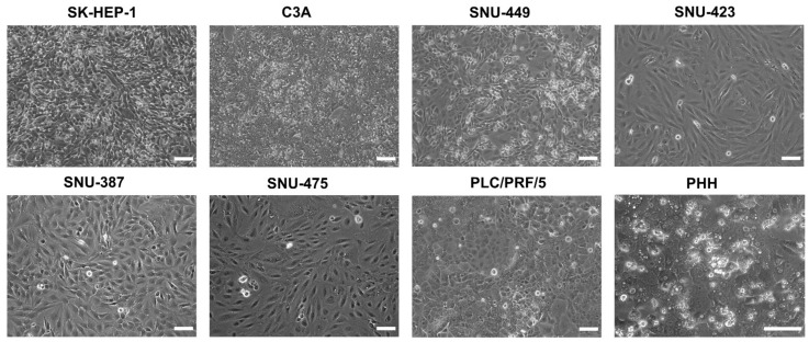
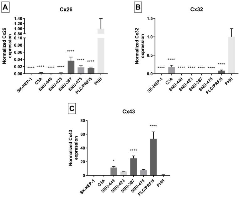
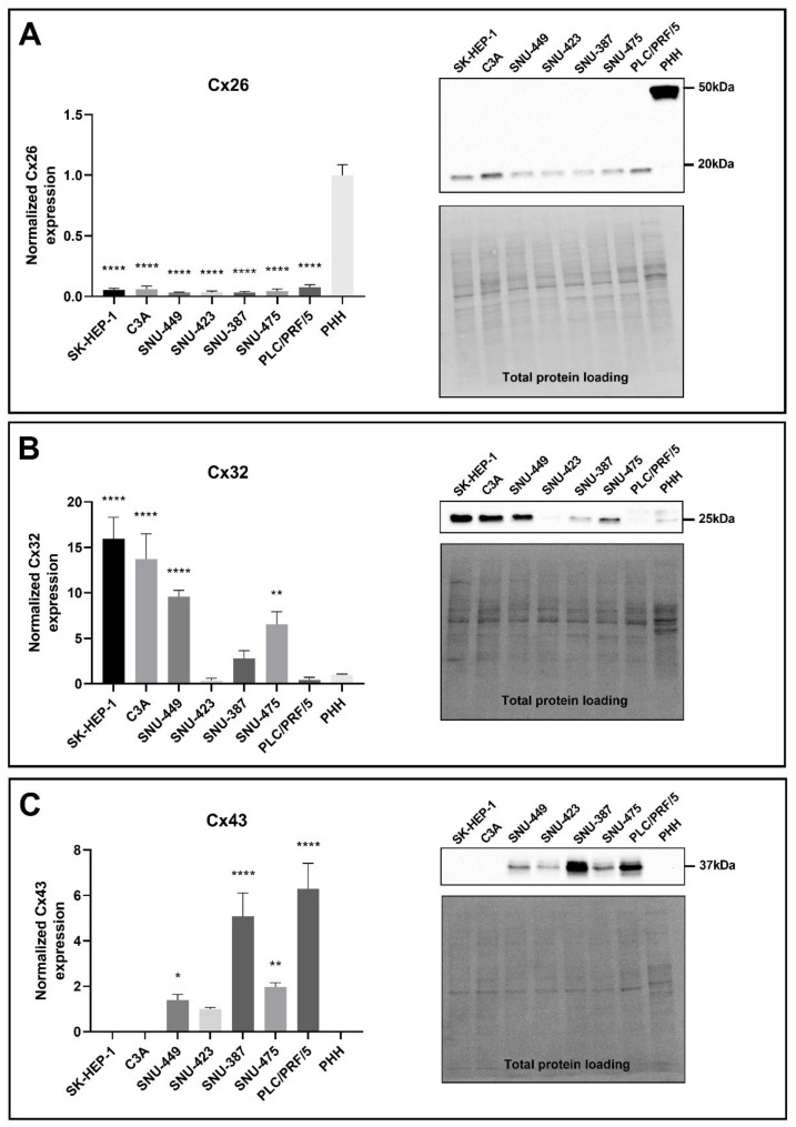
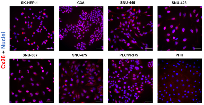
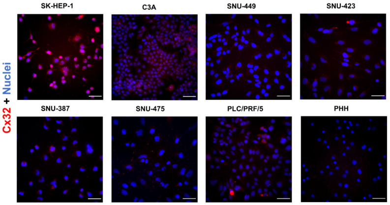
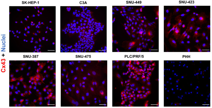
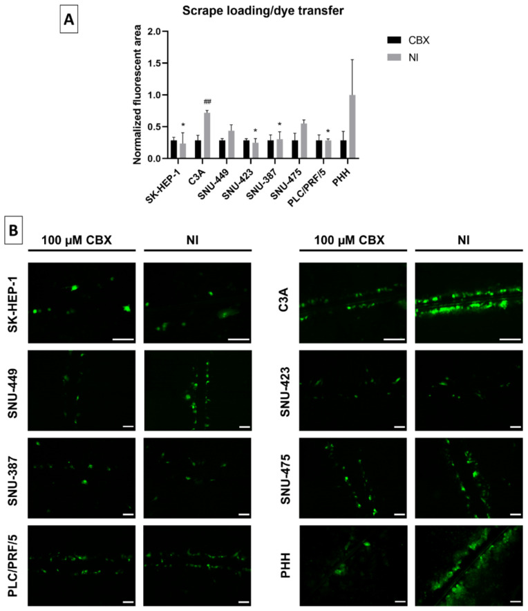
Liver cancer cell lines are frequently used in vitro tools to test candidate anti-cancer agents as well as to elucidate mechanisms of liver carcinogenesis. Among such mechanisms is cellular communication mediated by connexin-based gap junctions. The present study investigated changes in connexin expression and gap junction functionality in liver cancer in vitro. For this purpose, seven human liver cancer cell lines, as well as primary human hepatocytes, were subjected to connexin and gap junction analysis at the transcriptional, translational and activity level. Real-time quantitative reverse transcription polymerase chain reaction analysis showed enhanced expression of connexin43 in the majority of liver cancer cell lines at the expense of connexin32 and connexin26. Some of these changes were paralleled at the protein level, as evidenced by immunoblot analysis and in situ immunocytochemistry. Gap junctional intercellular communication, assessed by the scrape loading/dye transfer assay, was generally low in all liver cancer cell lines. Collectively, these results provide a full scenario of modifications in hepatocyte connexin production and gap junction activity in cultured liver cancer cell lines. The findings may be valuable for the selection of neoplastic hepatocytes for future mechanistic investigation and testing of anti-cancer drugs that target connexins and their channels.

Keywords: cell line; connexin; gap junction; in vitro; liver cancer.


Conflict of interest statement

The authors declare no conflict of interest.

Figures

Figure 1
Figure 1
Phase contrast images of liver cancer cell lines and primary human hepatocytes (PHH) at 100% confluence. All assays and extractions were performed when liver cancer cell lines reached 100% confluence. Phase contrast images were taken on a Nikon Eclipse Ti-S microscope (Nikon, Tokyo, Japan) with a 10× (liver cancer cell lines) or 20× objective (PHH). Scale bar = 100 µm.
Figure 2
Figure 2
Cx26 (A), Cx32 (B) and Cx43 (C) gene expression in liver cancer cell lines and primary human hepatocytes (PHH). Cancer cell lines were grown to 100% confluence, while PHH were used in suspension when total RNA was extracted (n = 1, N = 3). Subsequently, real-time quantitative reverse transcription polymerase chain reaction (RT-qPCR) analysis was performed. Relative alterations compared to PHH were calculated according to the Pfaffl method in qbase+ (Biogazelle, Gent, Belgium). Data are expressed as mean ± standard deviation with * p ≤ 0.05 and **** p ≤ 0.0001 compared to the PHH control.
Figure 3
Figure 3
Cx26 (A), Cx32 (B) and Cx43 (C) protein expression in liver cancer cell lines and primary human hepatocytes (PHH). Cancer cell lines (n = 1, N = 3) were grown to 100% confluence, while PHH were used in suspension for protein extraction. Immunoblotting and visualization were done with the Pierce™ ECL Western Blotting Substrate kit (Thermo Fisher Scientific, Waltham, MA, USA) on a ChemiDocTM MP imaging system. All signals were divided by their respective total protein loading signal and normalized by the sum of all data points in a replicate [42]. Unlike Cx43, which was not expressed by PHH, Cx26 and Cx32 are expressed relative to their expression in PHH. Data are expressed as mean ± standard deviation with * p ≤ 0.05, ** p ≤ 0.01 and **** p ≤ 0.0001 compared to the PHH control.
Figure 4
Figure 4
Cx26 protein localization in liver cancer cell lines and primary human hepatocytes (PHH). Cancer cell lines (n = 1, N = 2) were fixed during the exponential growth phase, while PHH were fixed at the final day of the sandwich cultivation period. All cells were immunostained for Cx26 (red) with nuclear counterstaining using 4′,6-diamidino-2-phenylindole (DAPI) (blue). Scale bar = 50 µm. Images were taken with a 40× objective. A representative image is shown.
Figure 5
Figure 5
Cx32 protein localization in liver cancer cell lines and primary human hepatocytes (PHH). Cancer cell lines (n = 1, N = 2) were fixed during the exponential growth phase, while PHH were fixed at the final day of the sandwich cultivation period. All cells were immunostained for Cx32 (red) with nuclear counterstaining using 4′,6-diamidino-2-phenylindole (DAPI) (blue). Scale bar = 50 µm. Images were taken with a 40× objective. A representative image is shown.
Figure 6
Figure 6
Cx43 protein localization in liver cancer cell lines and primary human hepatocytes (PHH). Cancer cell lines (n = 1, N = 2) were fixed during the exponential growth phase, while PHH were fixed at the final day of the sandwich cultivation period. All cells were immunostained for Cx43 (red) with nuclear counterstaining using 4′,6-diamidino-2-phenylindole (DAPI) (blue). Scale bar = 50 µm. Images were taken with a 40× objective. A representative image is shown.
Figure 7
Figure 7
Gap junctional intercellular communication (GJIC) in liver cancer cell lines. All cell lines (n = 1–2, N = 1 (multiple images per well)) were grown to 100% confluence when a scrape was made across the well in presence of Lucifer Yellow (LY). Cells were washed and subjected to fluorescence microscopy analysis. Images were taken with a 10× or 20× objective (B). GJIC was either not inhibited (NI) or inhibited by 100 µM carbenoxolone disodium salt (CBX) for 10 min. The fluorescent area was measured with ImageJ. Data were normalized to the inhibited control and expressed as a ratio to the mean NI fluorescent area of primary human hepatocytes (PHH). Data are expressed as mean ± standard deviation (A). Significant differences compared to NI PHH are indicated with * p ≤ 0.05, while differences to the respective CBX control are indicated with ## p ≤ 0.01. Scale bar = 100 µm.

Similar articles

Cited by

References

    1. Sung H., Ferlay J., Siegel R.L., Laversanne M., Soerjomataram I., Jemal A., Bray F. Global Cancer Statistics 2020: GLOBOCAN estimates of incidence and mortality worldwide for 36 cancers in 185 countries. CA Cancer J. Clin. 2021;71:209–249. doi: 10.3322/caac.21660. - DOI - PubMed
    1. Koumbi L. Dietary factors can protect against liver cancer development. World J. Hepatol. 2017;9:119–125. doi: 10.4254/wjh.v9.i3.119. - DOI - PMC - PubMed
    1. Sia D., Villanueva A., Friedman S.L., Llovet J.M. Liver cancer cell of origin, molecular class, and effects on patient prognosis. Gastroenterology. 2017;152:745–761. doi: 10.1053/j.gastro.2016.11.048. - DOI - PubMed
    1. Alqahtani A., Khan Z., Alloghbi A., Said Ahmed T.S., Ashraf M., Hammouda D.M. Hepatocellular carcinoma: Molecular mechanisms and targeted therapies. Medicina. 2019;55:526. doi: 10.3390/medicina55090526. - DOI - PMC - PubMed
    1. Collins S.D., Yuen G., Tu T., Budzinska M.A., Spring K., Bryant K., Shackel N.A. In vitro models of the liver: Disease modeling, drug discovery and clinical applications. In: Tirnitz-Parker J.E.E., editor. Hepatocellular Carcinoma. Codon Publications; Brisbane, Australia: 2019. - PubMed