. 2021 Nov 10;22(22):12189.
doi: 10.3390/ijms222212189.

The Effect of Tuberculosis Antimicrobials on the Immunometabolic Profiles of Primary Human Macrophages Stimulated with Mycobacterium tuberculosis

Affiliations

The Effect of Tuberculosis Antimicrobials on the Immunometabolic Profiles of Primary Human Macrophages Stimulated with Mycobacterium tuberculosis

Christina Cahill et al. Int J Mol Sci. .

Abstract

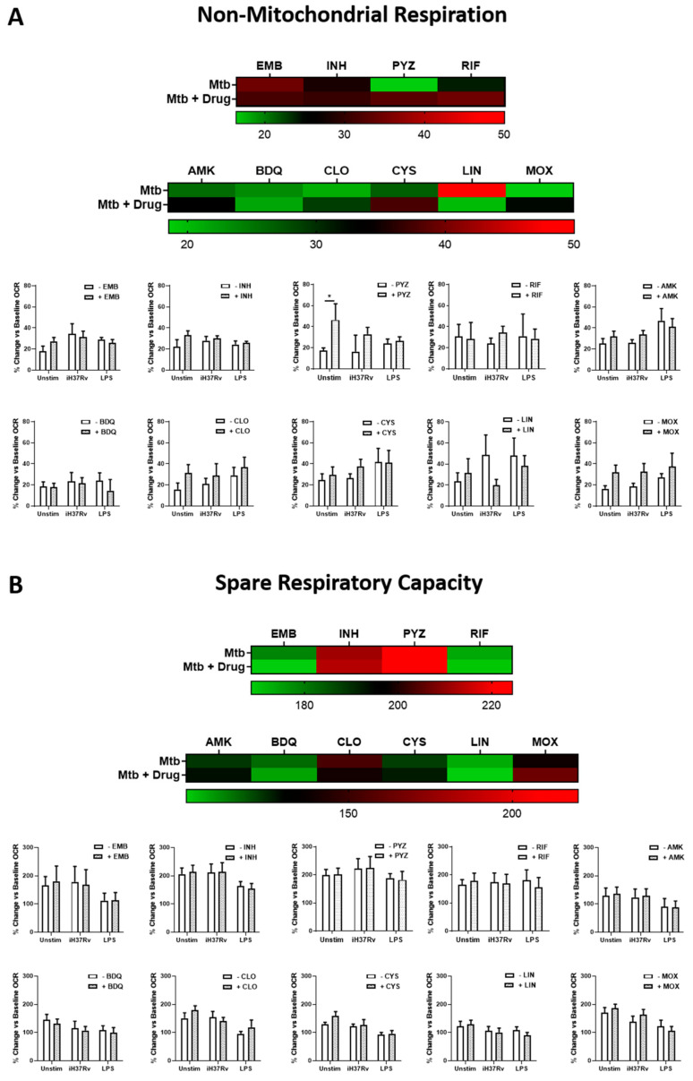
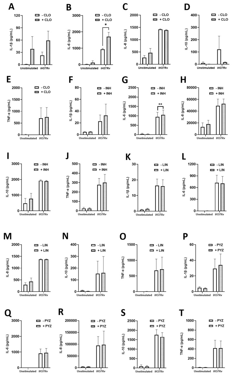
Tuberculosis (TB) remains a global health challenge. Patients with drug-sensitive and drug-resistant TB undergo long, arduous, and complex treatment regimens, often involving multiple antimicrobials. While these drugs were initially implemented based on their bactericidal effects, some studies show that TB antimicrobials can also directly affect cells of the immune system, altering their immune function. As use of these antimicrobials has been the mainstay of TB therapy for over fifty years now, it is more important than ever to understand how these antimicrobials affect key pathways of the immune system. One such central pathway, which underpins the immune response to a variety of infections, is immunometabolism, namely glycolysis and oxidative phosphorylation (OXPHOS). We hypothesise that in addition to their direct bactericidal effect on Mycobacterium tuberculosis (Mtb), current TB antimicrobials can modulate immunometabolic profiles and alter mitochondrial function in primary human macrophages. Human monocyte-derived macrophages (hMDMs) were differentiated from PBMCs isolated from healthy blood donors, and treated with four first-line and six second-line TB antimicrobials three hours post stimulation with either iH37Rv-Mtb or lipopolysaccharide (LPS). 24 h post stimulation, baseline metabolism and mitochondrial function were determined using the Seahorse Extracellular Flux Analyser. The effect of these antimicrobials on cytokine and chemokine production was also assayed using Meso Scale Discovery Multi-Array technology. We show that some of the TB antimicrobials tested can significantly alter OXPHOS and glycolysis in uninfected, iH37Rv-Mtb, and LPS-stimulated hMDMs. We also demonstrate how these antimicrobial-induced immunometabolic effects are linked with alterations in mitochondrial function. Our results show that TB antimicrobials, specifically clofazimine, can modify host immunometabolism and mitochondrial function. Moreover, clofazimine significantly increased the production of IL-6 in human macrophages that were stimulated with iH37Rv-Mtb. This provides further insight into the use of some of these TB antimicrobials as potential host-directed therapies in patients with early and active disease, which could help to inform TB treatment strategies in the future.

Keywords: antimicrobials; bioenergetics; drug-resistant tuberculosis; glycolysis; host-directed therapy; lipopolysaccharide; mitochondrial function; oxidative phosphorylation; tuberculosis.


Conflict of interest statement

The authors declare no conflict of interest.

Figures

Figure 1
Figure 1
Isoniazid, pyrazinamide, bedaquiline, clofazimine and linezolid increase glycolytic profiles in unstimulated, iH37Rv-Mtb-stimulated and LPS-stimulated hMDMs. (A) Illustrative heat map showing changes in ECAR in response to treatment with first-line (top) and second-line (bottom) TB antimicrobials. hMDMs, differentiated from PBMCs isolated from healthy blood donors, were treated with (B) ethambutol (5 µg/mL), (C) isoniazid (1 µg/mL), (D) pyrazinamide (2 µg/mL), (E) rifampicin (2 µg/mL), (F) amikacin (5 µg/mL), (G) bedaquiline (5 µg/mL), (H) clofazimine (2 µg/mL), (I) cycloserine (5 µg/mL), (J) linezolid (15 µg/mL) or (K) moxifloxacin (12.5 µg/mL) three hours post stimulation with either iH37Rv-Mtb or LPS (100 ng/mL). 24 h post stimulation, ECAR profiles, representing glycolysis, were determined utilising the Seahorse Extracellular Flux Analyser. (L) Summary of how the antimicrobials affect glycolytic profiles in unstimulated, iH37Rv-Mtb-stimulated and LPS-stimulated hMDMs. Bars denote mean ± SEM * p < 0.05 and ** p < 0.01 (Two-way repeated measures ANOVA with Sidak’s multiple comparisons test). Ethambutol (EMB, n = 7); isoniazid (INH, n = 8); pyrazinamide (PYZ, n = 7); rifampicin (RIF, n = 9); amikacin (AMK, n = 5); bedaquiline (BDQ, n = 6); clofazimine (CLO, n = 6); cycloserine (CYS, n = 4); linezolid (LIN, n = 8); moxifloxacin (MOX, n = 10).
Figure 2
Figure 2
Moxifloxacin and clofazimine reduces oxidative phosphorylation in unstimulated and iH37Rv-Mtb-stimulated hMDMs, respectively. (A) Illustrative heap map showing alterations in OCR in response to treatment with first-line (top) and second-line (bottom) TB antimicrobials. hMDMs, differentiated from PBMCs isolated from healthy blood donors, were treated with (B) ethambutol (5 µg/mL), (C) isoniazid (1 µg/mL), (D) pyrazinamide (2 µg/mL), (E) rifampicin (2 µg/mL), (F) amikacin (5 µg/mL), (G) bedaquiline (5 µg/mL), (H) clofazimine (2 µg/mL), (I) cycloserine (5 µg/mL), (J) linezolid (15 µg/mL) or (K) moxifloxacin (12.5 µg/mL) three hours post stimulation with either iH37Rv-Mtb or LPS (100 ng/mL). 24 h post stimulation, OCR profiles, representing oxidative phosphorylation, were determined utilising the Seahorse Extracellular Flux Analyser. (L) Summary of how the antimicrobials affect glycolytic profiles in unstimulated, and iH37Rv-Mtb-stimulated hMDMs. Bars denote mean ± SEM * p < 0.05 and ** p < 0.01 (Two-way repeated measures ANOVA with Sidak’s multiple comparisons test). Ethambutol (EMB, n = 7); isoniazid (INH, n = 8); pyrazinamide (PYZ, n = 7); rifampicin (RIF, n = 9); amikacin (AMK, n = 5); bedaquiline (BDQ, n = 6); clofazimine (CLO, n = 6); cycloserine (CYS, n = 4); linezolid (LIN, n = 8); moxifloxacin (MOX, n = 10).
Figure 3
Figure 3
Clofazimine treated hMDMs stimulated with iH37Rv-Mtb rely on glycolysis by promoting Warburg metabolism. hMDMs, differentiated from PBMCs isolated from healthy blood donors, were treated with (A) ethambutol (5 µg/mL), (B) isoniazid (1 µg/mL), (C) pyrazinamide (2 µg/mL), (D) rifampicin (2 µg/mL), (E) amikacin (5 µg/mL), (F) bedaquiline (5 µg/mL), (G) clofazimine (2 µg/mL), (H) cycloserine (5 µg/mL), (I) linezolid (15 µg/mL) or (J) moxifloxacin (12.5 µg/mL) three hours post stimulation with either iH37Rv-Mtb or LPS (100 ng/mL). At 24 h post stimulation, the ECAR:OCR ratio was generated to measure the reliance of one metabolic pathway over another. (K) Summary of how clofazimine affects the ECAR:OCR ratio in unstimulated and iH37Rv-Mtb-stimulated hMDMs. This immunometabolic shift as a result of clofazimine treatment is illustrated by a metabolic phenogram in (L) unstimulated and (M) Mtb-stimulated hMDMs. Bars denote mean ± SEM * p < 0.05 and ** p < 0.01 (Two-way repeated measures ANOVA with Sidak’s multiple comparisons test). Ethambutol (EMB, n = 7); isoniazid (INH, n = 8); pyrazinamide (PYZ, n = 7); rifampicin (RIF, n = 9); amikacin (AMK, n = 5); bedaquiline (BDQ, n = 6); clofazimine (CLO, n = 6); cycloserine (CYS, n = 4); linezolid (LIN, n = 8); moxifloxacin (MOX, n = 10).
Figure 4
Figure 4
TB antimicrobials do not affect spare respiratory capacity or non-mitochondrial respiration in iH37Rv-Mtb-stimulated and LPS-stimulated hMDMs. hMDMs, differentiated from PBMCs isolated from healthy blood donors, were treated with ethambutol (5 µg/mL), isoniazid (1 µg/mL), pyrazinamide (2 µg/mL), rifampicin (2 µg/mL), amikacin (5 µg/mL), bedaquiline (5 µg/mL), clofazimine (2 µg/mL), cycloserine (5 µg/mL), linezolid (15 µg/mL) or moxifloxacin (12.5 µg/mL) three hours post stimulation with either iH37Rv-Mtb or LPS (100 ng/mL). 24 h post stimulation, (A) non-mitochondrial respiration and (B) spare respiratory capacity were determined utilising the Seahorse Extracellular Flux Analyser. Bars denote mean ± SEM * p < 0.05 (Two-way repeated measures ANOVA with Sidak’s multiple comparisons test). Ethambutol (EMB, n = 7); isoniazid (INH, n = 6); pyrazinamide (PYZ, n = 7); rifampicin (RIF, n = 9); amikacin (AMK, n = 5); bedaquiline (BDQ, n = 6); clofazimine (CLO, n = 6); cycloserine (CYS, n = 4); linezolid (LIN, n = 8); moxifloxacin (MOX, n = 10).
Figure 5
Figure 5
Clofazimine reduces mitochondrial coupling efficiency and increases mitochondrial proton leak in iH37Rv-Mtb-stimulated hMDMs. hMDMs, differentiated from PBMCs isolated from healthy blood donors, were treated with ethambutol (5 µg/mL), isoniazid (1 µg/mL), pyrazinamide (2 µg/mL), rifampicin (2 µg/mL), amikacin (5 µg/mL), bedaquiline (5 µg/mL), clofazimine (2 µg/mL), cycloserine (5 µg/mL), linezolid (15 µg/mL) or moxifloxacin (12.5 µg/mL) three hours post stimulation with either iH37Rv-Mtb or LPS (100 ng/mL). 24 h post stimulation, (A) mitochondrial coupling efficiency and (B) mitochondrial proton leak were determined using the Seahorse Extracellular Flux Analyser. Bars denote mean ± SEM * p < 0.05 (Two-way repeated measures ANOVA with Sidak’s multiple comparisons test). Ethambutol (EMB, n = 7); isoniazid (INH, n = 8); pyrazinamide (PYZ, n = 7); rifampicin (RIF, n = 9); amikacin (AMK, n = 5); bedaquiline (BDQ, n = 6); clofazimine (CLO, n = 6); cycloserine (CYS, n = 4); linezolid (LIN, n = 8); moxifloxacin (MOX, n = 10).
Figure 6
Figure 6
Examining the effect of clofazimine, isoniazid, linezolid and pyrazinamide on protein levels of IL-1β, IL-6, IL-8, IL-10 and TNFα in hMDMs stimulated with iH37Rv-Mtb. hMDMs, differentiated from PBMCs isolated from healthy blood donors, were stimulated with iH37Rv Mtb for three hours, washed to remove unphagocytosed Mtb, and were treated with clofazimine (2 µg/mL), isoniazid (1 µg/mL), linezolid (15 µg/mL) or pyrazinamide (2 µg/mL). At 24 h post stimulation, protein levels of IL1β (A,F,K and P), IL-6 (B,G,L and Q), IL-8 (C,H,M and R), IL-10 (D,I,N and S) and TNFα (E,J,O and T) were quantified using Meso Scale Discovery Multi-Array technology. Bars denote mean ± SEM. * p < 0.05 (Two-way repeated measures ANOVA tests with Šídák’s multiple comparisons tests).
Figure 7
Figure 7
Assessing the effect of clofazimine, isoniazid, linezolid and pyrazinamide on protein levels of MCP-1, MCP-4, IP-10, MDC and MIP-1β in hMDMs stimulated with iH37Rv-Mtb. hMDMs, differentiated from PBMCs isolated from healthy blood donors, were stimulated with iH37Rv-Mtb for three hours, washed to remove unphagocytosed Mtb, and were treated with clofazimine (2 µg/mL), isoniazid (1 µg/mL), linezolid (15 µg/mL) or pyrazinamide (2 µg/mL). At 24 h post stimulation, protein levels of MCP-1 (A,F,K and P), MCP-4 (B,G,L and Q), IP-10 (C,H,M and R), MDC (D,I,N and S) and MIP-1β (E,J,O and T) were quantified using Meso Scale Discovery Multi-Array technology. Bars denote mean ± SEM. * p < 0.05 (Two-way repeated measures ANOVA tests with Šídák’s multiple comparisons tests).

References

    1. Mitini-Nkhoma S.C., Chimbayo E.T., Mzinza D.T., Mhango D.V., Chirambo A.P., Mandalasi C., Lakudzala A.E., Tembo D.L., Jambo K.C., Mwandumba H.C. Something Old, Something New: Ion Channel Blockers as Potential Anti-Tuberculosis Agents. Front. Immunol. 2021;12:2203. doi: 10.3389/fimmu.2021.665785. - DOI - PMC - PubMed
    1. Krug S., Parveen S., Bishai W.R. Host-Directed Therapies: Modulating Inflammation to Treat Tuberculosis. Front. Immunol. 2021;12:1247. doi: 10.3389/fimmu.2021.660916. - DOI - PMC - PubMed
    1. Hayford F.E.A., Dolman R.C., Blaauw R., Nienaber A., Smuts C.M., Malan L., Ricci C. The effects of anti-inflammatory agents as host-directed adjunct treatment of tuberculosis in humans: A systematic review and meta-analysis. Respir. Res. 2020;21:223. doi: 10.1186/s12931-020-01488-9. - DOI - PMC - PubMed
    1. Cox D.J., Coleman A.M., Gogan K.M., Phelan J.J., Maoldomhnaigh C.Ó., Dunne P.J., Basdeo S.A., Keane J. Inhibiting Histone Deacetylases in Human Macrophages Promotes Glycolysis, IL-1β, and T Helper Cell Responses to Mycobacterium tuberculosis. Front. Immunol. 2020;11:1609. doi: 10.3389/fimmu.2020.01609. - DOI - PMC - PubMed
    1. Cahill C., O’Connell F., Gogan K.M., Cox D.J., Basdeo S.A., O’Sullivan J., Gordon S.V., Keane J., Phelan J.J. The Iron Chelator Desferrioxamine Increases the Efficacy of Bedaquiline in Primary Human Macrophages Infected with BCG. Int. J. Mol. Sci. 2021;22:2938. doi: 10.3390/ijms22062938. - DOI - PMC - PubMed

MeSH terms