

3D Cell Culture Systems: Tumor Application, Advantages, and Disadvantages
- PMID: 34830082
- PMCID: PMC8618305
- DOI: 10.3390/ijms222212200
3D Cell Culture Systems: Tumor Application, Advantages, and Disadvantages
Abstract
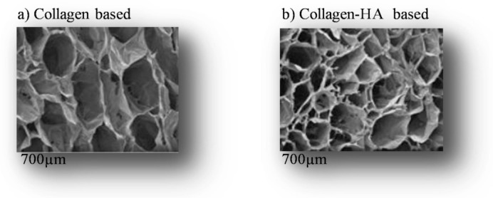
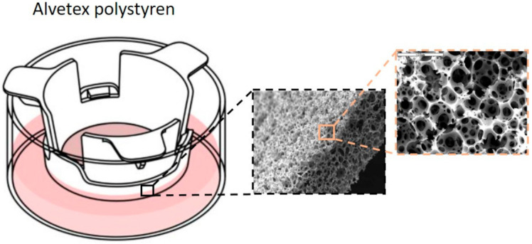
The traditional two-dimensional (2D) in vitro cell culture system (on a flat support) has long been used in cancer research. However, this system cannot be fully translated into clinical trials to ideally represent physiological conditions. This culture cannot mimic the natural tumor microenvironment due to the lack of cellular communication (cell-cell) and interaction (cell-cell and cell-matrix). To overcome these limitations, three-dimensional (3D) culture systems are increasingly developed in research and have become essential for tumor research, tissue engineering, and basic biology research. 3D culture has received much attention in the field of biomedicine due to its ability to mimic tissue structure and function. The 3D matrix presents a highly dynamic framework where its components are deposited, degraded, or modified to delineate functions and provide a platform where cells attach to perform their specific functions, including adhesion, proliferation, communication, and apoptosis. So far, various types of models belong to this culture: either the culture based on natural or synthetic adherent matrices used to design 3D scaffolds as biomaterials to form a 3D matrix or based on non-adherent and/or matrix-free matrices to form the spheroids. In this review, we first summarize a comparison between 2D and 3D cultures. Then, we focus on the different components of the natural extracellular matrix that can be used as supports in 3D culture. Then we detail different types of natural supports such as matrigel, hydrogels, hard supports, and different synthetic strategies of 3D matrices such as lyophilization, electrospiding, stereolithography, microfluid by citing the advantages and disadvantages of each of them. Finally, we summarize the different methods of generating normal and tumor spheroids, citing their respective advantages and disadvantages in order to obtain an ideal 3D model (matrix) that retains the following characteristics: better biocompatibility, good mechanical properties corresponding to the tumor tissue, degradability, controllable microstructure and chemical components like the tumor tissue, favorable nutrient exchange and easy separation of the cells from the matrix.
Keywords: extracellular matrix; hydrogel; spheroids; three-dimensional (3D) culture model; tissue engineering.
Conflict of interest statement
The authors have no financial disclosures to make or conflict to report.
Figures


References
Publication types
MeSH terms
LinkOut - more resources
Full Text Sources
Medical
