. 2021 Nov 15;22(22):12332.
doi: 10.3390/ijms222212332.

NTRK Gene Fusion Detection in Atypical Spitz Tumors

Affiliations

NTRK Gene Fusion Detection in Atypical Spitz Tumors

Rocco Cappellesso et al. Int J Mol Sci. .

Abstract

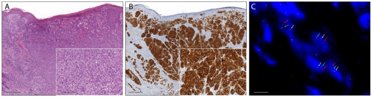
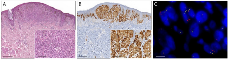
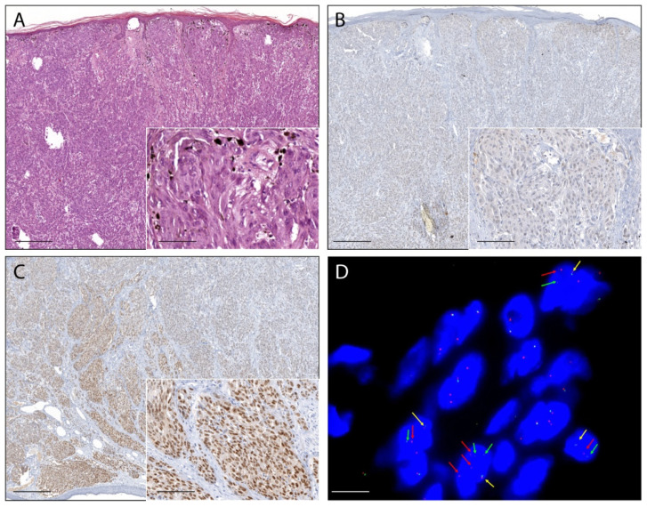
Atypical Spitz tumors (AST) deviate from stereotypical Spitz nevi for one or more atypical features and are now regarded as an intermediate category of melanocytic tumors with uncertain malignant potential. Activating NTRK1/NTRK3 fusions elicit oncogenic events in Spitz lesions and are targetable with kinase inhibitors. However, their prevalence among ASTs and the optimal approach for their detection is yet to be determined. A series of 180 ASTs were screened with pan-TRK immunohistochemistry and the presence of NTRK fusions was confirmed using FISH, two different RNA-based NGS panels for solid tumors, and a specific real time RT-PCR panel. Overall, 26 ASTs showed pan-TRK immunostaining. NTRK1 fusions were detected in 15 of these cases showing cytoplasmic immunoreaction, whereas NTRK3 was detected in one case showing nuclear immunoreaction. Molecular tests resulted all positive in only two ASTs (included the NTRK3 translocated), RNA-based NGS and real time RT-PCR were both positive in three cases, and FISH and real time RT-PCR in another two cases. In seven ASTs NTRK1 fusions were detected only by FISH and in two cases only by real time RT-PCR. The frequency of NTRK fusions in ASTs is 9%, with a clear prevalence of NTRK1 compared to NTRK3 alterations. Pan-TRK immunohistochemistry is an excellent screening test. Confirmation of NTRK fusions may require the use of different molecular techniques.

Keywords: NTRK; NTRK1; NTRK3; atypical spitz tumor; pan-TRK.


Conflict of interest statement

DM has received honoraria for professional services and consultancy for Bayer HealthCare Pharmaceuticals Inc. (Whippany, NJ, USA). The remaining authors declare that they have no conflict of interest.

Figures

Figure 1
Figure 1
Photomicrographs of an atypical Spitz tumor harboring anNTRK1 fusion. (A): The lesion was an exophytic nodule with epidermal collarette, flat lower border, and focal epidermal hyperplasia with filigree-like rete ridges (H&E staining, magnification 40×, scale bar: 200 µm). Inset: lobulated nests of epithelioid melanocytes with distinct cell borders and quite pleomorphic nuclei (H&E staining, magnification 200×, scale bar: 40 µm). (B): Melanocytes showed strong and diffuse cytoplasmic immunoreaction (pan-TRK immunostaining, magnification 40×, scale bar: 200 µm). Inset: pan-TRK cytoplasmatic immunoreaction (pan-TRK immunostaining, magnification 200×, scale bar: 40 µm). (C): Break-apart FISH analysis confirmed NTRK1 rearrangement showing the typical pattern: one fusion signal (yellow arrows) and split signals 3′ (red arrows) and 5′ (green arrows) (ZytoLight SPEC NTRK1 Dual Color Break Apart Probe, original magnification 1000×, scale bar: 5 µm).
Figure 2
Figure 2
Photomicrographs of an atypical Spitz tumor harboring an NTRK1 fusion. (A): The lesion was dome-shaped with epidermal hyperplasia (H&E staining, magnification 40×, scale bar: 200 µm). Inset: lobulated nests of epithelioid melanocytes with distinct cell borders and quite pleomorphic nuclei showing a tendency towards maturation that became exaggerated at the bottom of the lesion (H&E staining, magnification 200×, scale bar: 40 µm). (B): Melanocytes showed strong and diffuse cytoplasmic immunoreaction (pan-TRK immunostaining, magnification 40×, scale bar 200 µm). Inset: pan-TRK cytoplasmatic immunoreaction (pan-TRK immunostaining, magnification 200×, scale bar 40 µm). (C): Break-apart FISH analysis confirmed NTRK1 rearrangement showing the atypical pattern: one fusion signal (yellow arrows) and a single orange signal 3′ (red arrows) (ZytoLight SPEC NTRK1 Dual Color Break Apart Probe, original magnification 1000×, scale bar 5 µm).
Figure 3
Figure 3
Photomicrographs of an atypical Spitz tumor harboring an NTRK3 fusion. (A): The lesion was an exophytic nodule with epidermal collarette, flat lower border, and widespread epidermal hyperplasia with long and thin filigree-like rete ridges enveloping superficial nests. (H&E staining, magnification 40×, scale bar 200 µm). Inset: melanocytes were predominantly elongated, with ground-glass cytoplasm, and arranged in fascicles (H&E staining, magnification 200×, scale bar 40 µm). (B): In some areas, the melanocytes lacked immunoreaction (pan-TRK immunostaining, magnification 40×, scale bar: 200 µm). Inset: melanocytes without immunoreaction (pan-TRK immunostaining, magnification 200×, scale bar 40 µm); (C): Most of the melanocytes showed strong nuclear immunoreaction (pan-TRK immunostaining, magnification 40×, scale bar: 200 µm). Inset: melanocytes with nuclear immunoreaction (pan-TRK immunostaining, original magnification 200×, scale bar: 40 µm). (D): Break-apart FISH analysis confirmed NTRK3 rearrangement showing the typical pattern: one fusion signal (yellow arrows) and split signals 3′ (red arrows) and 5′ (green arrows) (ZytoLight SPEC NTRK3 Dual Color Break Apart Probe, original magnification 1000×, scale bar 5 µm).

Similar articles

Cited by

References

    1. Massi D., De Giorgi V., Mandalà M. The complex management of atypical Spitz tumours. Pathology. 2016;48:132–141. doi: 10.1016/j.pathol.2015.12.003. - DOI - PubMed
    1. Ferrara G., Cavicchini S., Corradin M.T. Hypopigmented atypical Spitzoid neoplasms (atypical Spitz nevi, atypical Spitz tumors, Spitzoid melanoma): A clinicopathological update. Derm. Pr. Concept. 2015;30:45–52. doi: 10.5826/dpc.0501a06. - DOI - PMC - PubMed
    1. Gerami P., Busam K., Cochran A., Cook M.G., Duncan L.M., Elder D.E., Fullen D.R., Guitart J., LeBoit P.E., Mihm M.C., Jr., et al. Histomorphologic assessment and interobserver diagnostic reproducibility of atypical spitzoid melanocytic neoplasms with long-term follow-up. Am. J. Surg. Pathol. 2014;38:934–940. doi: 10.1097/PAS.0000000000000198. - DOI - PubMed
    1. Cerroni L., Barnhill R., Elder D., Gottlieb G., Heenan P., Kutzner H., LeBoit P.E., Mihm M., Jr., Rosai J., Kerl H. Melanocytic tumors of uncertain malignant potential: Results of a tutorial held at the XXIX Symposium of the International Society of Dermatopathology in Graz, October 2008. Am. J. Surg. Pathol. 2010;34:314–326. doi: 10.1097/PAS.0b013e3181cf7fa0. - DOI - PubMed
    1. Gill M., Renwick N., Silvers D.N., Celebi J.T. Lack of BRAF mutations in Spitz nevi. J. Investig. Dermatol. 2004;122:1325–1326. doi: 10.1111/j.0022-202X.2004.22530.x. - DOI - PubMed

MeSH terms