

The Emerging Role of Decellularized Plant-Based Scaffolds as a New Biomaterial
- PMID: 34830229
- PMCID: PMC8625747
- DOI: 10.3390/ijms222212347
The Emerging Role of Decellularized Plant-Based Scaffolds as a New Biomaterial
Abstract
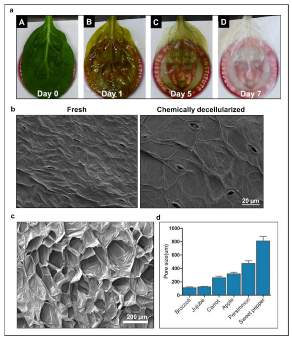
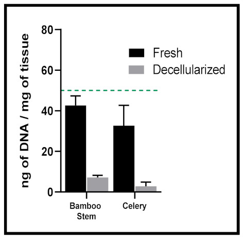
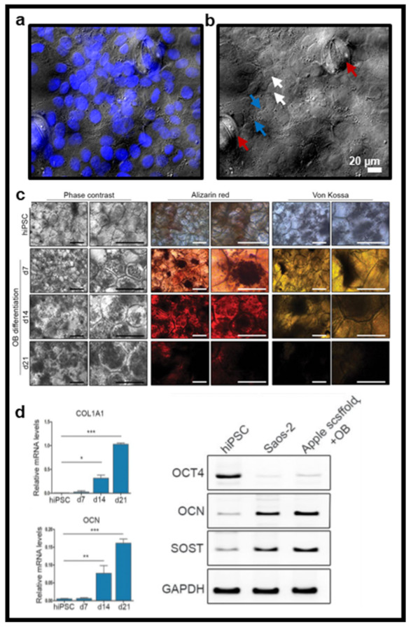
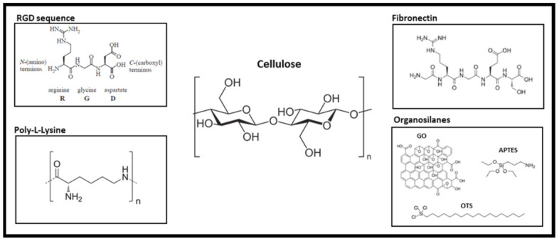
The decellularization of plant-based biomaterials to generate tissue-engineered substitutes or in vitro cellular models has significantly increased in recent years. These vegetal tissues can be sourced from plant leaves and stems or fruits and vegetables, making them a low-cost, accessible, and sustainable resource from which to generate three-dimensional scaffolds. Each construct is distinct, representing a wide range of architectural and mechanical properties as well as innate vasculature networks. Based on the rapid rise in interest, this review aims to detail the current state of the art and presents the future challenges and perspectives of these unique biomaterials. First, we consider the different existing decellularization techniques, including chemical, detergent-free, enzymatic, and supercritical fluid approaches that are used to generate such scaffolds and examine how these protocols can be selected based on plant cellularity. We next examine strategies for cell seeding onto the plant-derived constructs and the importance of the different functionalization methods used to assist in cell adhesion and promote cell viability. Finally, we discuss how their structural features, such as inherent vasculature, porosity, morphology, and mechanical properties (i.e., stiffness, elasticity, etc.) position plant-based scaffolds as a unique biomaterial and drive their use for specific downstream applications. The main challenges in the field are presented throughout the discussion, and future directions are proposed to help improve the development and use of vegetal constructs in biomedical research.
Keywords: biomaterial; cellulose; decellularization; plant-based scaffolds; tissue engineering.
Conflict of interest statement
Authors A.F.H., J.L. and F.Z. are authors on patent WO 2020/264385 A1.
Figures
Similar articles
-
Supercritical carbon dioxide decellularization of plant material to generate 3D biocompatible scaffolds.Sci Rep. 2021 Feb 11;11(1):3643. doi: 10.1038/s41598-021-83250-9. Sci Rep. 2021. PMID: 33574461 Free PMC article.
-
Decellularized orthopaedic tissue-engineered grafts: biomaterial scaffolds synthesised by therapeutic cells.Biomater Sci. 2018 Oct 24;6(11):2798-2811. doi: 10.1039/c8bm00772a. Biomater Sci. 2018. PMID: 30229775 Review.
-
Recent advances in plant-derived polysaccharide scaffolds in tissue engineering: A review.Int J Biol Macromol. 2024 Oct;277(Pt 1):133830. doi: 10.1016/j.ijbiomac.2024.133830. Epub 2024 Jul 11. Int J Biol Macromol. 2024. PMID: 39002914 Review.
-
Decellularized natural 3D cellulose scaffold derived from Borassus flabellifer (Linn.) as extracellular matrix for tissue engineering applications.Carbohydr Polym. 2021 Nov 15;272:118494. doi: 10.1016/j.carbpol.2021.118494. Epub 2021 Jul 29. Carbohydr Polym. 2021. PMID: 34420749
-
Optimizing decellularization protocols for human thyroid tissues: a step towards tissue engineering and transplantation.Biomed Mater. 2024 Jun 20;19(4). doi: 10.1088/1748-605X/ad565e. Biomed Mater. 2024. PMID: 38857607
Cited by
-
Fibrin and Marine-Derived Agaroses for the Generation of Human Bioartificial Tissues: An Ex Vivo and In Vivo Study.Mar Drugs. 2023 Mar 17;21(3):187. doi: 10.3390/md21030187. Mar Drugs. 2023. PMID: 36976236 Free PMC article.
-
Plant-Based Decellularization: A Novel Approach for Perfusion-Compatible Tissue Engineering Structures.J Microbiol Biotechnol. 2024 May 28;34(5):1003-1016. doi: 10.4014/jmb.2401.01024. Epub 2024 Feb 29. J Microbiol Biotechnol. 2024. PMID: 38563106 Free PMC article. Review.
-
Cultivation of bovine lipid chunks on Aloe vera scaffolds.NPJ Sci Food. 2025 Feb 25;9(1):26. doi: 10.1038/s41538-025-00391-1. NPJ Sci Food. 2025. PMID: 40000634 Free PMC article.
-
Current advances in the development of microRNA-integrated tissue engineering strategies: a cornerstone of regenerative medicine.Front Bioeng Biotechnol. 2024 Oct 16;12:1484151. doi: 10.3389/fbioe.2024.1484151. eCollection 2024. Front Bioeng Biotechnol. 2024. PMID: 39479296 Free PMC article. Review.
-
Effect of mechanical forces on cellular response to radiation.Radiother Oncol. 2022 Nov;176:187-198. doi: 10.1016/j.radonc.2022.10.006. Epub 2022 Oct 10. Radiother Oncol. 2022. PMID: 36228760 Free PMC article. Review.
References
-
- Gilpin A., Yang Y. Decellularization Strategies for Regenerative Medicine: From Processing Techniques to Applications. [(accessed on 8 May 2019)]. Available online: https://www.hindawi.com/journals/bmri/2017/9831534/ - PMC - PubMed
-
- O’Brien F.J. Biomaterials & Scaffolds for Tissue Engineering. Mater. Today. 2011;14:88–95. doi: 10.1016/S1369-7021(11)70058-X. - DOI
Publication types
MeSH terms
Substances
LinkOut - more resources
Full Text Sources
Other Literature Sources
