. 2021 Nov 22;22(22):12604.
doi: 10.3390/ijms222212604.

Connexin46 Expression Enhances Cancer Stem Cell and Epithelial-to-Mesenchymal Transition Characteristics of Human Breast Cancer MCF-7 Cells

Affiliations

Connexin46 Expression Enhances Cancer Stem Cell and Epithelial-to-Mesenchymal Transition Characteristics of Human Breast Cancer MCF-7 Cells

Rodrigo A Acuña et al. Int J Mol Sci. .

Abstract

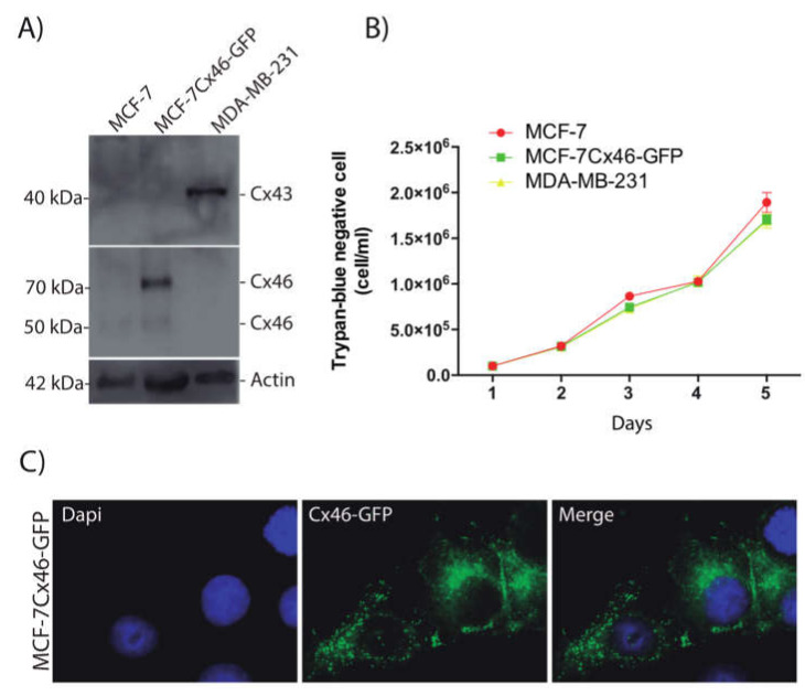
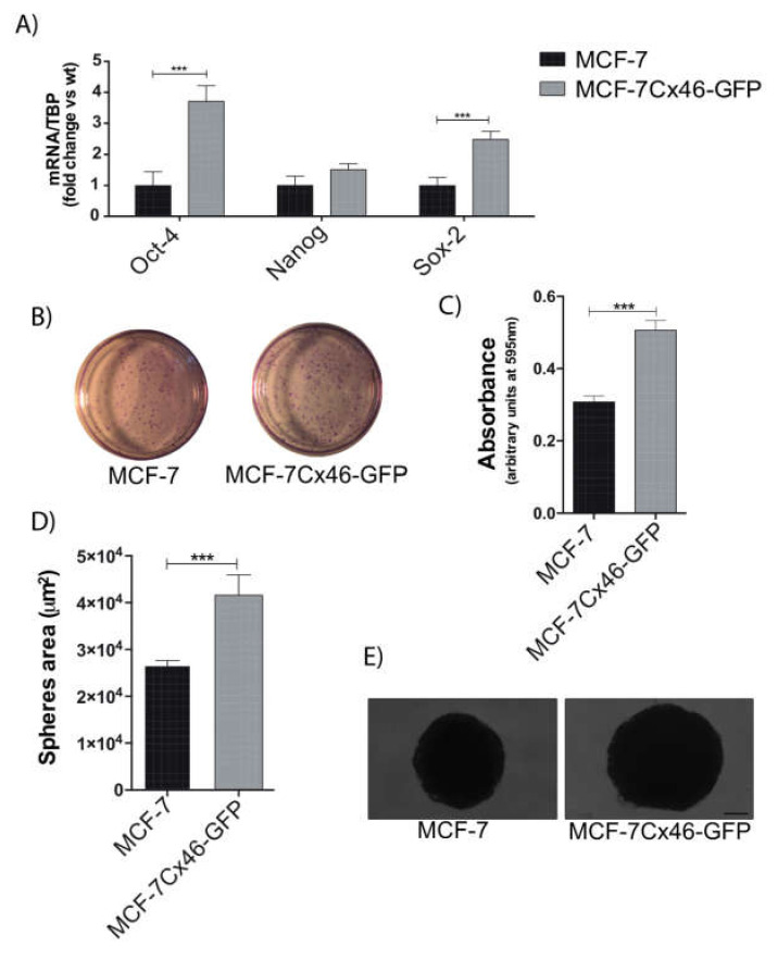
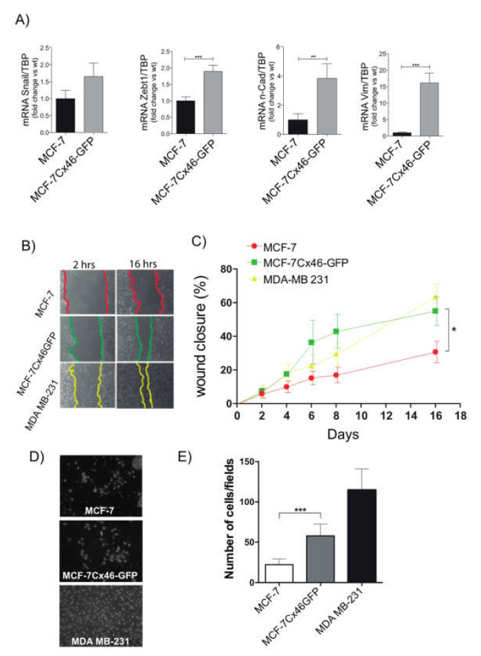
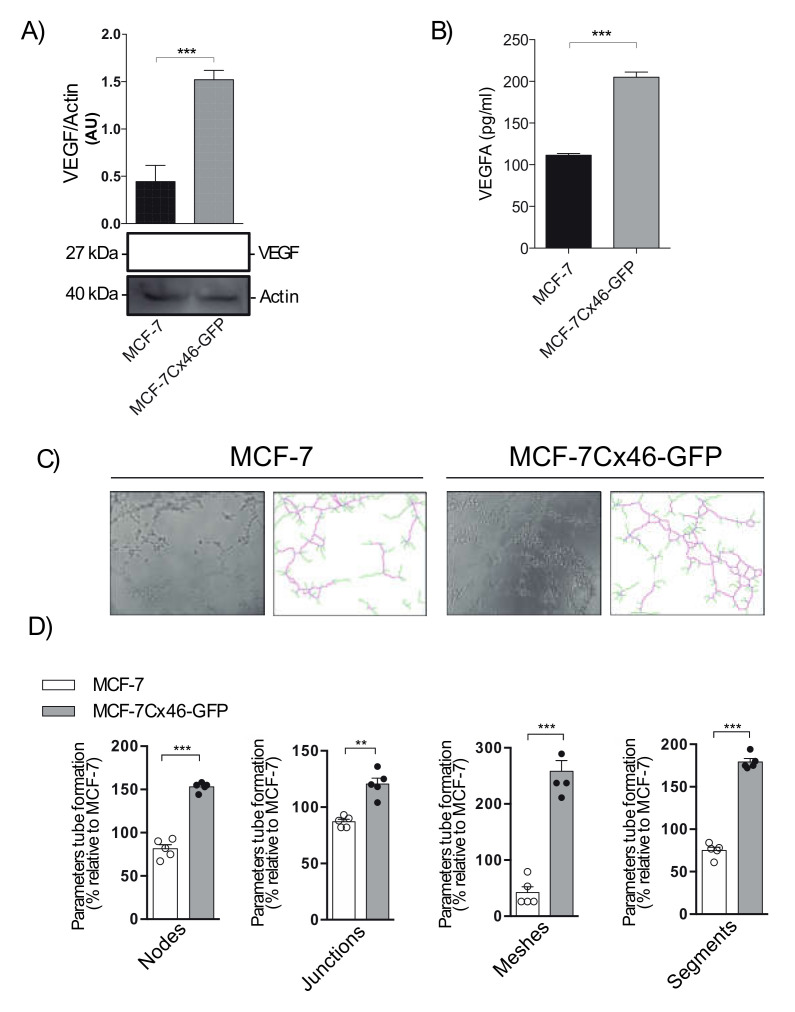
Connexins (Cxs) are a family of proteins that form two different types of ion channels: hemichannels and gap junction channels. These channels participate in cellular communication, enabling them to share information and act as a synchronized syncytium. This cellular communication has been considered a strong tumor suppressor, but it is now recognized that some type of Cxs can be pro-tumorigenic. For example, Cx46 expression is increased in human breast cancer samples and correlates with cancer stem cell (CSC) characteristics in human glioma. Thus, we explored whether Cx46 and glioma cells, can set up CSC and epithelial-to-mesenchymal transition (EMT) properties in a breast cancer cell line. To this end, we transfected MCF-7 cells with Cx46 attached to a green fluorescent protein (Cx46GFP), and we determined how its expression orchestrates both the gene-expression and functional changes associated with CSC and EMT. We observed that Cx46GFP increased Sox2, Nanog, and OCT4 mRNA levels associated with a high capacity to form monoclonal colonies and tumorspheres. Similarly, Cx46GFP increased the mRNA levels of n-cadherin, Vimentin, Snail and Zeb1 to a higher migratory and invasive capacity. Furthermore, Cx46GFP transfected in MCF-7 cells induced the release of higher amounts of VEGF, which promoted angiogenesis in HUVEC cells. We demonstrated for the first time that Cx46 modulates CSC and EMT properties in breast cancer cells and thus could be relevant in the design of future cancer therapies.

Keywords: Connexin46; breast cancer; cancer stem cells; epithelial-to-mesenchymal transition; stemness.


Conflict of interest statement

The authors declare no conflict of interest, financial or otherwise.

Figures

Figure 1
Figure 1
Connexin 46-GFP overexpressed in MCF-7 cells does not modify the rate of cell division. MCF-7 breast cancer cells were transfected with the human connexin Cx-46 gene inserted in a pcmV6-AC-GFP plasmid and selected by neomycin resistance. (A) Representative images of three independent Western blots of MCF-7, MCF-7Cx46-GFP, and MDAMB-231 cell lysates; immunoblots were incubated with anti Cx46, and anti-beta Actin (Santa Cruz Biotechnology; 1:2500). Protein bands were detected using Immobilon Forte Western horseradish protein (HRP) substrate and visualized with a LI-COR CDigit System. (B) A trypan blue dye exclusion test determined the number of viable cells. MCF-7, MCF7Cx46-GFP, and MDAMB-231 were seeded, and the trypan blue negative cells were counted in a Neubauer chamber. The cells were counted every day until the fifth day. (C) Representative fluorescence images of MCF-7Cx46-GFP cell nuclei were visualized with Dapi (left), and Cx46 was visualized with the GFP tag in the C-terminal portion of Cx46 (middle). Images were obtained using a Nikon Eclipse Ti-U inverted microscope. Data are presented as the mean of three independent experiments +/− SEM.
Figure 2
Figure 2
Cx46 increases the expression of CSC-related genes and CSC stemness. (A) Cancer stem cell-related genes were evaluated by measuring the mRNA levels of Oct4, Nanog, and Sox2 genes by qRT-PCR in MCF-7 and MCF-7Cx46-GFP breast cancer cells; data represent the mean of three independent experiments +/− SEM. (B) Clonogenic potential of MCF-7 and MCF-7Cx46-GFP breast cancer cells. Representative images of clonogenic assay were fixed and stained with crystal violet solution. (C) Cells were dissolved with lysis buffer and absorbance was measured at 595 nm (arbitrary units at 595 nm). The results represent the means of three independent experiments +/− SEM. (D) The graph represents the comparison of sphere diameters between MCF-7 and MCF-7Cx46-GFP before 3 weeks of culture. (E) Representative image of 3-week sphere morphology of MCF-7 and MCF-7Cx46-GFP cells; scale bar 100 μm. Data are presented as the mean of three independent experiments +/− SEM. *** p < 0.001.
Figure 3
Figure 3
Cx46-GFP increases the expression of EMT-related genes and EMT functional characteristics. (A) The EMT genes Snail, Zebt1, N-Caderin and Vimentin of MCF-7 and MCF-7Cx46-GFP were determined by qRT-PCR. The graph represents the means of three independent experiments. (B) In vitro scratch assays determined differences in breast cancer cell migration. The image in (B) was recorded every 2 h and photos were obtained at the indicated time points using a 10× objective on a Nikon inverted microscope, recorded and measured using the NIS-Element software. The figure shows images at 2 and 16 h for each cell line, and the color line shows the gap area in the three different cell lines evaluated. (C) Graphical representations of percentage of wound closure area of the three different experiments represented in B. The closing percentage was calculated at different times until the 16 h period was complete. Data represent the means of three independent experiments +/− SEM. * denotes p < 0.05, ** p < 0.01 and *** p < 0.001. (D) Representative images of fields in a Transwell migration assay of MCF-7 (up), MCF-7Cx46-GFP (center), and MDAMB-231 (bottom); cells that crossed the gel matrix were fixed and stained with Dapi. (E) Ten different fields were photographed, and the number of cells was counted and graphed. Data are presented as the means of three independent experiments +/− SEM.
Figure 4
Figure 4
Cx46 increases the expression of angiogenic factors. (A) Representative Western blot of VEGF expression in MCF-7 and MCF-7Cx46-GFP and densitometry quantification of three independent Western blots of VEGF protein expression. (B) Graphical representation of VEGF release in conditioned supernatants media evaluated using a Luminex MAGPIX system. (C) Representative images of tubule structure formation in HUVEC cell incubated with conditioned medium from MCF-7 (left) and MCF-7Cx46-GFP (right) cells. (D) Representative graph quantification of different tube formation parameters (junctions, nodes, meshes, and branches) in HUVEC cells incubated with conditioned medium from MCF-7 and MCF-7Cx46-GFP. After 16 h, the tube formation was examined by phase contrast and the images were captured using an Olympus U-RFL-T camera. Junctions, nodes, meshes, and branches were analyzed using the ImageJ Angiogenesis Analyzer software. Data are presented as the means of three independent experiments +/− SEM. ** p < 0.01 and *** p < 0.001.

References

    1. Sáez J.C., Berthoud V.M., Brañes M.C., Martínez A.D., Beyer E.C. Plasma Membrane Channels Formed by Connexins: Their Regulation and Functions. Physiol. Rev. 2003;83:1359–1400. doi: 10.1152/physrev.00007.2003. - DOI - PubMed
    1. Retamal M.A., Reyes E.P., García I.E., Pinto B., Martínez A.D., González C. Diseases associated with leaky hemichannels. Front. Cell. Neurosci. 2015;9:267. doi: 10.3389/fncel.2015.00267. - DOI - PMC - PubMed
    1. Yamasaki H. Role of cell-cell communication in tumor suppression. Immunol. Ser. 1990;51:245–266. - PubMed
    1. Aasen T., Leithe E., Graham S.V., Kameritsch P., Mayán M.D., Mesnil M., Pogoda K., Tabernero A. Connexins in cancer: Bridging the gap to the clinic. Oncogene. 2019;38:4429–4451. doi: 10.1038/s41388-019-0741-6. - DOI - PMC - PubMed
    1. Paul D.L., Ebihara L., Takemoto L.J., Swenson K.I., Goodenough D.A. Connexin46, a novel lens gap junction protein, induces voltage-gated currents in nonjunctional plasma membrane of Xenopus oocytes. J. Cell Biol. 1991;115:1077–1089. doi: 10.1083/jcb.115.4.1077. - DOI - PMC - PubMed

MeSH terms