

MRI Findings in Hip in Juvenile Idiopathic Arthritis
- PMID: 34830537
- PMCID: PMC8625848
- DOI: 10.3390/jcm10225252
MRI Findings in Hip in Juvenile Idiopathic Arthritis
Abstract
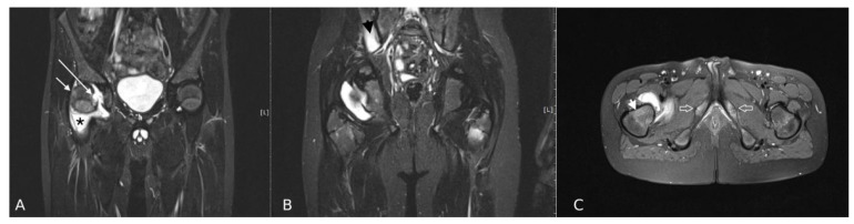
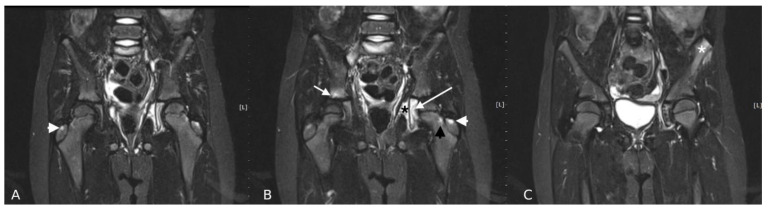
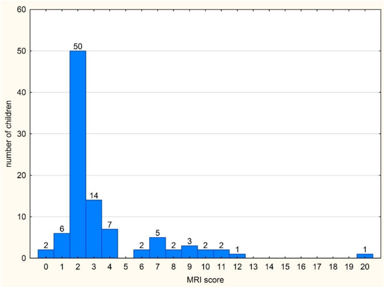
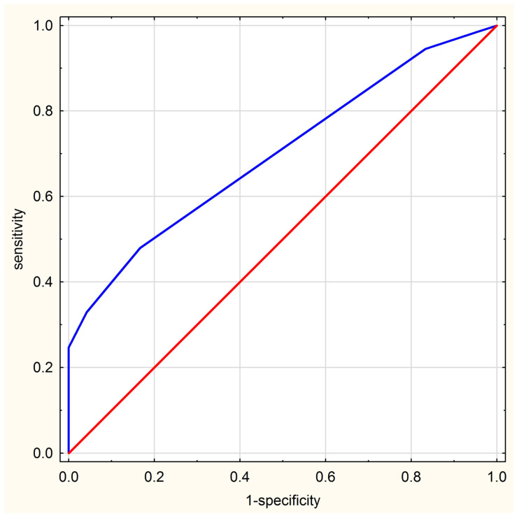
The aim of this study was to evaluate if magnetic resonance imaging allows hip arthritis in JIA to be differentiated from hip arthralgia of unknown etiology in juveniles clinically suspected for hip arthritis. This was a retrospective observational study which included 97 children with clinically suspected hip arthritis. Each hip was assessed and scored in MRI for signs of active and destructive inflammatory lesions and developmental lesions. MRI findings between JIA-confirmed patients and without final diagnosis of JIA were compared and the MRI summarized score was calculated, as the sum of scorings of all 24 hip lesions in an individual patient (i.a., effusion, synovitis, bone marrow edema, enthesitis). MRI showed at least one lesion in the majority of patients (95 patients; 98%). Effusion was the most common feature, followed by bone marrow oedema and synovitis. All lesions were more common in patients with a final diagnosis of JIA, especially synovitis and enthesitis (p = 0.037 and p = 0.047). The MRI summarized score was significantly higher in the JIA group than the non-JIA group: 3 (2-5) vs. 2 (2-2), respectively, p = 0.002. Using a cut-off score of 6, the MRI summarized score showed 25% sensitivity and 100% specificity indicating a good ability in discriminating hip arthritis during JIA from non-JIA patients. MRI allows hip arthritis in JIA to be differentiated from hip arthralgia of unknown etiology with good specificity, thus, may be helpful in confirming the diagnosis of JIA.
Keywords: arthralgia; arthritis; hip; juvenile; magnetic resonance imaging.
Conflict of interest statement
The authors declare no conflict of interest.
Figures
References
-
- Lovell D.J. Juvenile idiopathic arthritis: Clinical features. In: Klippel J.H., Stone J.H., Crofford L.J., White P.F., editors. Primer on the Rheumatic Diseases. Springer; New York, NY, USA: 2008. pp. 142–148.
-
- El-Azeem M.I.A., Taha H.A., El-Sherif A.M. Role of MRI in evaluation of hip joint involvement in juvenile idiopathic arthritis. Egypt. Rheumatol. 2012;34:75–82. doi: 10.1016/j.ejr.2012.03.001. - DOI
LinkOut - more resources
Full Text Sources
