Review
. 2021 Nov 12;13(22):5650.
doi: 10.3390/cancers13225650.

Microtubule Targeting Agents in Disease: Classic Drugs, Novel Roles

Affiliations
Review

Microtubule Targeting Agents in Disease: Classic Drugs, Novel Roles

Linda Wordeman et al. Cancers (Basel). .

Abstract

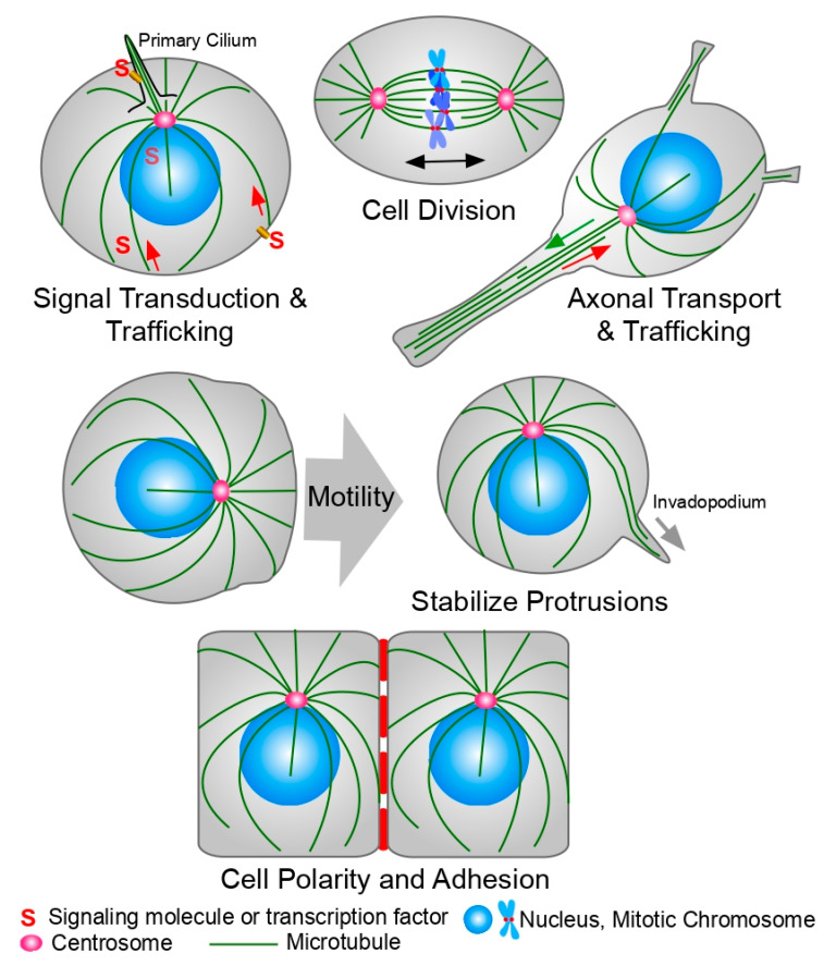
Microtubule-targeting agents (MTAs) represent one of the most successful first-line therapies prescribed for cancer treatment. They interfere with microtubule (MT) dynamics by either stabilizing or destabilizing MTs, and in culture, they are believed to kill cells via apoptosis after eliciting mitotic arrest, among other mechanisms. This classical view of MTA therapies persisted for many years. However, the limited success of drugs specifically targeting mitotic proteins, and the slow growing rate of most human tumors forces a reevaluation of the mechanism of action of MTAs. Studies from the last decade suggest that the killing efficiency of MTAs arises from a combination of interphase and mitotic effects. Moreover, MTs have also been implicated in other therapeutically relevant activities, such as decreasing angiogenesis, blocking cell migration, reducing metastasis, and activating innate immunity to promote proinflammatory responses. Two key problems associated with MTA therapy are acquired drug resistance and systemic toxicity. Accordingly, novel and effective MTAs are being designed with an eye toward reducing toxicity without compromising efficacy or promoting resistance. Here, we will review the mechanism of action of MTAs, the signaling pathways they affect, their impact on cancer and other illnesses, and the promising new therapeutic applications of these classic drugs.

Keywords: cancer; microtubule-targeting agent (MTA); microtubules (MTs); migration; pathogen; tauopathies; vascular formation.


Conflict of interest statement

The authors declare no conflict of interest.

Figures

Figure 1
Figure 1
Microtubules (MTs) functions in the cell. MTs play roles in a variety of cellular functions. Top row, left: MTs are involved in the transport of a variety of molecules, vesicles, and organelles inside the cell, affecting signaling transduction pathways. They are also essential in the formation and maintenance of the primary cilium, a signaling hub for the cell. Top row, center: MTs form the mitotic spindle, the machinery in charge of chromosome segregation during mitosis. Top row, right: MTs form the tracks used by molecular motors to transport neurotransmitters through the axon to the synaptic cleft. Center row: Proper organization of the MT cytoskeleton and centrosome localization is important to stabilize the migrating leading edge, for cell motility and for the formation of the invadopodium, a cellular extension of the cell membrane required for invasion of adjacent tissues. Bottom row: MTs are also important during the adhesion and the basal-to-lumen cell polarity establishment of epithelial cells. They also play an important role during the formation and turnover of focal adhesions. See the text for details.
Figure 2
Figure 2
Microtubule-targeting agents (MTAs) biding sites on the tubulin dimer. The αβ-tubulin dimer is shown in gray and light brown with the α subunit on the left and the β subunit on the right. Colored representative structures of the ligands are located on their binding sites. Color code: blue, Pironetin; yellow, Gatorbulin; cyan, colchicine; red, taxane; orange, vinca; magenta, laulimalide/peloruside; green, Maytansine. See text for details.

Similar articles

Cited by

References

    1. Goodson H.V., Jonasson E.M. Microtubules and Microtubule-Associated Proteins. Cold Spring Harb. Perspect. Biol. 2018;10:a022608. doi: 10.1101/cshperspect.a022608. - DOI - PMC - PubMed
    1. Findeisen P., Mühlhausen S., Dempewolf S., Hertzog J., Zietlow A., Carlomagno T., Kollmar M. Six Subgroups and Extensive Recent Duplications Characterize the Evolution of the Eukaryotic Tubulin Protein Family. Genome Biol. Evol. 2014;6:2274–2288. doi: 10.1093/gbe/evu187. - DOI - PMC - PubMed
    1. Chaaban S., Brouhard G.J. A Microtubule Bestiary: Structural Diversity in Tubulin Polymers. Mol. Biol. Cell. 2017;28:2924–2931. doi: 10.1091/mbc.e16-05-0271. - DOI - PMC - PubMed
    1. Mitchison T.J. Localization of an Exchangeable GTP Binding Site at the plus End of Microtubules. Science. 1993;261:1044–1047. doi: 10.1126/science.8102497. - DOI - PubMed
    1. Mitchison T., Kirschner M. Dynamic Instability of Microtubule Growth. Nature. 1984;312:237–242. doi: 10.1038/312237a0. - DOI - PubMed

LinkOut - more resources