Review
. 2021 Nov 12;13(22):5666.
doi: 10.3390/cancers13225666.

The Classification of Myeloproliferative Neoplasms: Rationale, Historical Background and Future Perspectives with Focus on Unclassifiable Cases

Affiliations
Review

The Classification of Myeloproliferative Neoplasms: Rationale, Historical Background and Future Perspectives with Focus on Unclassifiable Cases

Marco Pizzi et al. Cancers (Basel). .

Abstract

Myeloproliferative neoplasms (MPNs) are a heterogeneous group of clonal hematopoietic stem cell disorders, characterized by increased proliferation of one or more myeloid lineages in the bone marrow. The classification and diagnostic criteria of MPNs have undergone relevant changes over the years, reflecting the increased awareness on these conditions and a better understanding of their biological and clinical-pathological features. The current World Health Organization (WHO) Classification acknowledges four main sub-groups of MPNs: (i) Chronic Myeloid Leukemia; (ii) classical Philadelphia-negative MPNs (Polycythemia Vera; Essential Thrombocythemia; Primary Myelofibrosis); (iii) non-classical Philadelphia-negative MPNs (Chronic Neutrophilic Leukemia; Chronic Eosinophilic Leukemia); and (iv) MPNs, unclassifiable (MPN-U). The latter are currently defined as MPNs with clinical-pathological findings not fulfilling the diagnostic criteria for any other entity. The MPN-U spectrum traditionally encompasses early phase MPNs, terminal (i.e., advanced fibrotic) MPNs, and cases associated with inflammatory or neoplastic disorders that obscure the clinical-histological picture. Several lines of evidence and clinical practice suggest the existence of additional myeloid neoplasms that may expand the spectrum of MPN-U. To gain insight into such disorders, this review addresses the history of MPN classification, the evolution of their diagnostic criteria and the complex clinical-pathological and biological features of MPN-U.

Keywords: MPN-U; Myeloproliferative neoplasms; WHO Classification; myeloid disorders.


Conflict of interest statement

The Authors declare no conflict of interest.

Figures
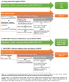
Figure 1
Figure 1
Diagnostic flow chart for patients with suspected MPN.
Figure 2
Figure 2
Representative histological features of case #1. BM biopsy shows cellularity within normal limits (a; H/E, 20×) with accumulation of hematopoiesis in central marrow spaces and marginalization of the adipose tissue close to the bony trabeculae. Maturation of erythroid and myeloid lineages is preserved (b; H/E, 200×), but scattered, large megakaryocytes are noted (b, inset). Reticulin network is unremarkable (c, Gomori silver stain, 200×).
Figure 3
Figure 3
Representative histological features of case #2, #3 and #4. BM biopsy from case #2 (ac) shows a hypercellular marrow with panmyelosis and loose clustering of megakaryocytes (a; H/E, 200×), featuring giant, hyperlobulated forms (b; H/E, 400×); reticulin network is unremarkable (c; Gomori silver stain, 200×). BM biopsy from case #3 (df) depicts hypercellularity with panmyelosis and loose to denser clustering of megakaryocytes (d; H/E, 200×), which range in morphology from small/hypolobulated to large/hyperlobulated (e, H/E, 400×); a minor increase in reticulin network, consistent with MF-1 grade is observed (f; Gomori silver stain, 200×). BM biopsy from case #4 (gi) features panmyelosis and loose clustering of megakaryocytes with small to giant, hyperlobulated forms and naked nuclei (g; H/E, 200×); megaloblastoid features of erythropoiesis are best appreciated with Giemsa stain (h; 400×), whereas Gomori stain is unremarkable (i; 200×).
Figure 4
Figure 4
Representative histological features of case #5 and #6. BM biopsy from case #5 (ac) shows a hypocellular marrow (a; H/E, 40×), featuring, at least focally, a mild decrease in myeloid-to-erythroid ratio without maturation defects (b; H/E, 400×), and scattered MPN-like, giant hyperlobulated megakaryocytes (c; H/E, 400×). BM biopsy from case #6 (df) depicts hypercellularity with increased myeloid-to-erythroid ratio, myeloid left shifting and clustering of megakaryocytes with hyperlobated forms and maturation defects (d; Giemsa, 400×; e; H/E 400×); a well-represented CD14+ monocyte population is present (f; 200×).
Figure 5
Figure 5
Representative histological features of case #7. BM biopsy from case #7 shows panmyelosis with normal myeloid-to-erythroid ratio, mildly left shifted granulopoiesis and increased, mature-looking megakaryocytes (a; H/E, 200×) with unremarkable reticulin network (b; Gomori silver stain, 200×) and a minor increase in CD34+ blasts (c; 200×).

Similar articles

Cited by

References

    1. Swerdlow S.H., Campo E., Harris N.L., Jaffe E.S., Pileri S.A., Stein H., Thiele J. WHO Classification of Tumours of Haematopoietic and Lymphoid Tissues. IARC Press; Lyon, France: 2017.
    1. Anderson L.A., McMullin M.F. Epidemiology of MPN: What Do We Know? Curr. Hematol. Malign. Rep. 2014;9:340–349. doi: 10.1007/s11899-014-0228-z. - DOI - PubMed
    1. Szuber N., Mudireddy M., Nicolosi M., Penna D., Vallapureddy R.R., Lasho T.L., Finke C., Begna K.H., Elliott M.A., Hook C.C., et al. 3023 Mayo Clinic Patients with Myeloproliferative Neoplasms: Risk-Stratified Comparison of Survival and Outcomes Data Among Disease Subgroups. Mayo Clin. Proc. 2019;94:599–610. doi: 10.1016/j.mayocp.2018.08.022. - DOI - PubMed
    1. Tefferi A. The history of myeloproliferative disorders: Before and after Dameshek. Leukemia. 2007;22:3–13. doi: 10.1038/sj.leu.2404946. - DOI - PubMed
    1. Dameshek W. Some speculations on the myeloproliferative syndromes. Blood. 1951;6:372–375. doi: 10.1182/blood.V6.4.372.372. - DOI - PubMed