Review
. 2021 Nov 12;13(22):5678.
doi: 10.3390/cancers13225678.

Impact of Chromatin Dynamics and DNA Repair on Genomic Stability and Treatment Resistance in Pediatric High-Grade Gliomas

Affiliations
Review

Impact of Chromatin Dynamics and DNA Repair on Genomic Stability and Treatment Resistance in Pediatric High-Grade Gliomas

Lia Pinto et al. Cancers (Basel). .

Abstract

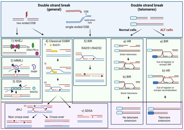
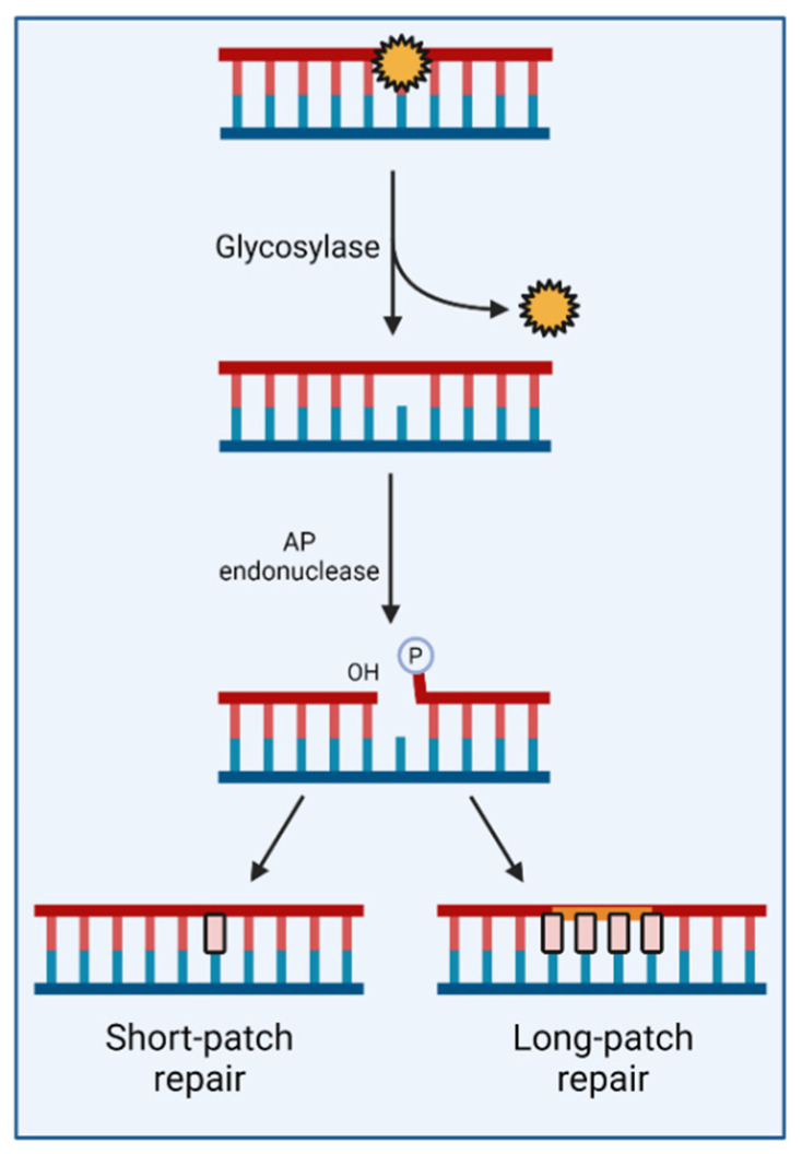
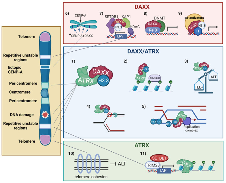
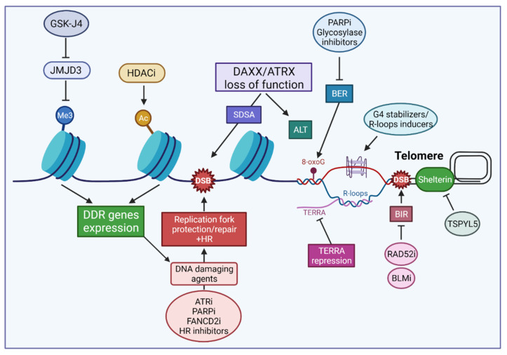
Despite their low incidence, pediatric high-grade gliomas (pHGGs), including diffuse intrinsic pontine gliomas (DIPGs), are the leading cause of mortality in pediatric neuro-oncology. Recurrent, mutually exclusive mutations affecting K27 (K27M) and G34 (G34R/V) in the N-terminal tail of histones H3.3 and H3.1 act as key biological drivers of pHGGs. Notably, mutations in H3.3 are frequently associated with mutations affecting ATRX and DAXX, which encode a chaperone complex that deposits H3.3 into heterochromatic regions, including telomeres. The K27M and G34R/V mutations lead to distinct epigenetic reprogramming, telomere maintenance mechanisms, and oncogenesis scenarios, resulting in distinct subgroups of patients characterized by differences in tumor localization, clinical outcome, as well as concurrent epigenetic and genetic alterations. Contrasting with our understanding of the molecular biology of pHGGs, there has been little improvement in the treatment of pHGGs, with the current mainstays of therapy-genotoxic chemotherapy and ionizing radiation (IR)-facing the development of tumor resistance driven by complex DNA repair pathways. Chromatin and nucleosome dynamics constitute important modulators of the DNA damage response (DDR). Here, we summarize the major DNA repair pathways that contribute to resistance to current DNA damaging agent-based therapeutic strategies and describe the telomere maintenance mechanisms encountered in pHGGs. We then review the functions of H3.3 and its chaperones in chromatin dynamics and DNA repair, as well as examining the impact of their mutation/alteration on these processes. Finally, we discuss potential strategies targeting DNA repair and epigenetic mechanisms as well as telomere maintenance mechanisms, to improve the treatment of pHGGs.

Keywords: (peri)centromere; ATRX; DAXX; DNA repair; alternative lengthening of telomere (ALT); chemoresistance; chromatin dynamics; genomic instability; pediatric high-grade gliomas; synthetic lethality; telomerase; telomere; variant H3.3 histone.


Conflict of interest statement

The authors declare no conflict of interest.

Figures

Figure 1
Figure 1
Schematics of the major DSB repair pathways. In NHEJ, the broken ends are bound by the Ku heterodimer, leading to the recruitment and activation of DNA-dependent protein kinase catalytic subunit (DNA-PKcs) and subsequent juxtaposition of the ends, followed by an eventual processing step—for instance, in the case of “dirty” ends produced by IR [33]—to generate ligatable ends. Central to HR, SSA and MMEJ is a DNA end resection step [34] that generates 3′-ended, single-stranded DNA (ssDNA) tails. In MMEJ, a pathway that is promoted by poly(ADP-ribose) Polymerase 1 (PARP1) acting in part to prevent Ku binding to DSBs [35], end-resection exposes short regions of complementary sequences (microhomologies) that facilitate end-bridging before processing the non-homologous flaps by the XPF/ERCC1 endonuclease (arrows), filling-in DNA synthesis and ligation. In SSA, homologous sequences exposed by long ssDNA strands are annealed by the RAD52 recombinase [36] to generate an intermediate with non-homologous flaps that are clipped by XPF/ERCC1, allowing filling-in DNA synthesis and ligation [37]. In DSBR, The RAD51 recombinase acts on long resected tails to assemble nucleoprotein filaments (i) that invade the homologous donor sequence to generate a displacement loop (D-loop) (ii) and prime repair DNA synthesis at the invading end, templated by the homologous sequence (iii) [31]. DSB repair can then proceed via classical DSBR (also called gene conversion) or synthesis-dependent strand annealing (SDSA). In DSBR, capture of the second end by the displaced strand of the D-loop is associated with extended DNA repair synthesis and the formation of a double Holliday junction (dHJ) (iv) whose resolution/dissolution can lead to non-crossovers or crossovers between the sister chromatids. On the other hand, SDSA involves limited DNA synthesis as well as the displacement of the invading strand from the donor sequence and its annealing to the second, unengaged resected end, thereby preventing dHJ formation and crossover products. Single-ended DSBs are formed when the replication fork collapses upon an encounter with, e.g., SSBs or trapped protein-DNA complexes. The repair of seDSBs can occur through HR or BIR mechanisms mediated by the RAD51 or RAD52 recombinase. BIR is initiated by strand invasion to form a D-loop and progresses via D-loop migration. Of note, recent studies suggest that RAD51-dependent BIR can also take place to repair two-ended DSBs in mammals, as previously shown in yeast [38,39]. BIR mechanisms and HR (DSBR and SDSA) can also take place at telomeres. In cells with intact ATRX/DAXX, the model of Lovejoy et al. [40] proposes that telomere cohesion promoted by ATRX fosters “in-register” HR/BIR using the sister telomere, while loss of ATRX promotes “out-of-register” or ectopic HR/BIR, resulting in a net increase in telomere length, as observed in ALT cells. The authors further propose that disruption of a DAXX-dependent function of ATRX promotes BIR over other pathways [40]. See text for details. For the sake of clarity, only a few of the DNA repair factors involved in DSB repair have been indicated. Created with BioRender.
Figure 2
Figure 2
Salient features of the base excision pathway. BER is initiated by a DNA glycosylase (e.g., OGG1, NTH1, NEIL1-3, UDG, MPG) that recognizes specific types of base damage and mediates the excision of the damaged base, creating an apurinic/apyrimidinic (AP) site. Cleavage of the phosphodiester backbone by AP endonuclease 1 (APE1) then generates an SSB intermediate with a 3′-OH and 5′-deoxyribose phosphate (5′dRP) residue which is processed to allow nucleotide replacement by repair DNA synthesis in steps that can involve the replacement of either a single nucleotide (short patch BER) or several nucleotides (long patch BER). Of note, certain DNA glycosylases are bifunctional, possessing an AP-lyase activity that can process the AP site. BER mediates the repair of the major lesions induced by TMZ (N7-methylguanine and N3-methyladenine). It also provides the major mechanism for the removal of oxidative damage lesions. The repair of SSBs (not illustrated here), which involves their recognition by PARP1, is considered a subpathway of BER. Created with BioRender.
Figure 3
Figure 3
Functions of the ATRX/DAXX histone H3.3 chaperone complex and mutually independent functions DAXX and ATRX in chromatin dynamics, DNA repair, and genomic stability. Central Panel: The ATRX/DAXX complex deposits H3.3 at heterochromatic loci with underlying repetitive DNA elements, including telomeres and (peri)centromeres (1). In cells undergoing global demethylation, ATRX/DAXX recruits the SUV39H1 histone methyltransferase to foster the heterochromatinization of specific tandem repetitive elements (2). ATRX/ADXX also prevents ALT development (3) and replication stress associated with the formation of secondary structures in repetitive DNA sequences (4). Finally, ATRX/DAXX-mediated deposition of H3.3 promotes D-loop stabilization and repair synthesis during DSB repair by HR (5). Other DNA repair functions are detailed in the text. Top panel: ATRX-independent functions for DAXX include the ectopic deposition of over-expressed CENP-A (6) and roles as a transcriptional activator or repressor depending on the context (7–9). In association with SETDB1, KAP1, and HDAC1, DAXX is responsible for the silencing of ERVs (7). DAXX recruitment to RelB-regulated genes contributes to their transcriptional silencing mediated by the DNA methyltransferase DNMT1 (8). DAXX may also exert transcriptional repression roles through interaction with HDACs (not illustrated). DAXX can also serve as a transcriptional co-activator in certain situations, such as during heat-induced activation of stress response genes (9). On its part, ATRX maintains cohesion between sister telomeres (10) and exerts a KAP1/TRIM28- and SETDB1-mediated repression of IAP retrotransposons by reinforcing the heterochromatin signature at these sites (11). See text for a full description of the DNA repair functions of H3.3/ATRX/DAXX. Created with BioRender.
Figure 4
Figure 4
Therapeutic strategies targeting DNA repair and telomere dynamics in pHGGs. Illustrated are current or proposed strategies affecting the expression of DNA repair genes through the targeting of epigenetic modifiers operating at marks impacted by the oncogenic histone H3.1/H3.3 mutations, or exploiting the replication stress inherent to pHGG cells, alone or in combination with DNA damaging agents. Other potential targets include components of the SDSA pathway, which is the favored HR DSB repair pathway upon loss of ATRX/DAXX. Also illustrated are strategies targeting telomeric DNA repair and telomere maintenance mechanisms. See text for details. Created with BioRender.

References

    1. Juratli T.A., Qin N., Cahill D.P., Filbin M.G. Molecular pathogenesis and therapeutic implications in pediatric high-grade gliomas. Pharmacol. Ther. 2018;182:70–79. doi: 10.1016/j.pharmthera.2017.08.006. - DOI - PubMed
    1. Hoffman L.M., Veldhuijzen van Zanten S.E.M., Colditz N., Baugh J., Chaney B., Hoffmann M., Lane A., Fuller C., Miles L., Hawkins C., et al. Clinical, Radiologic, Pathologic, and Molecular Characteristics of Long-Term Survivors of Diffuse Intrinsic Pontine Glioma (DIPG): A Collaborative Report From the International and European Society for Pediatric Oncology DIPG Registries. J. Clin. Oncol. 2018;36:1963–1972. doi: 10.1200/JCO.2017.75.9308. - DOI - PMC - PubMed
    1. Mackay A., Burford A., Carvalho D., Izquierdo E., Fazal-Salom J., Taylor K.R., Bjerke L., Clarke M., Vinci M., Nandhabalan M., et al. Integrated Molecular Meta-Analysis of 1000 Pediatric High-Grade and Diffuse Intrinsic Pontine Glioma. Cancer Cell. 2017;32:520–537.e5. doi: 10.1016/j.ccell.2017.08.017. - DOI - PMC - PubMed
    1. Schwartzentruber J., Korshunov A., Liu X.Y., Jones D.T., Pfaff E., Jacob K., Sturm D., Fontebasso A.M., Quang D.A., Tonjes M., et al. Driver mutations in histone H3.3 and chromatin remodelling genes in paediatric glioblastoma. Nature. 2012;482:226–231. doi: 10.1038/nature10833. - DOI - PubMed
    1. Sturm D., Witt H., Hovestadt V., Khuong-Quang D.A., Jones D.T., Konermann C., Pfaff E., Tonjes M., Sill M., Bender S., et al. Hotspot mutations in H3F3A and IDH1 define distinct epigenetic and biological subgroups of glioblastoma. Cancer Cell. 2012;22:425–437. doi: 10.1016/j.ccr.2012.08.024. - DOI - PubMed

LinkOut - more resources