Review
. 2021 Nov 13;13(22):5681.
doi: 10.3390/cancers13225681.

Biological Significance and Targeting of the FGFR Axis in Cancer

Affiliations
Review

Biological Significance and Targeting of the FGFR Axis in Cancer

Athina-Myrto Chioni et al. Cancers (Basel). .

Abstract

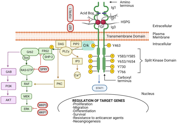
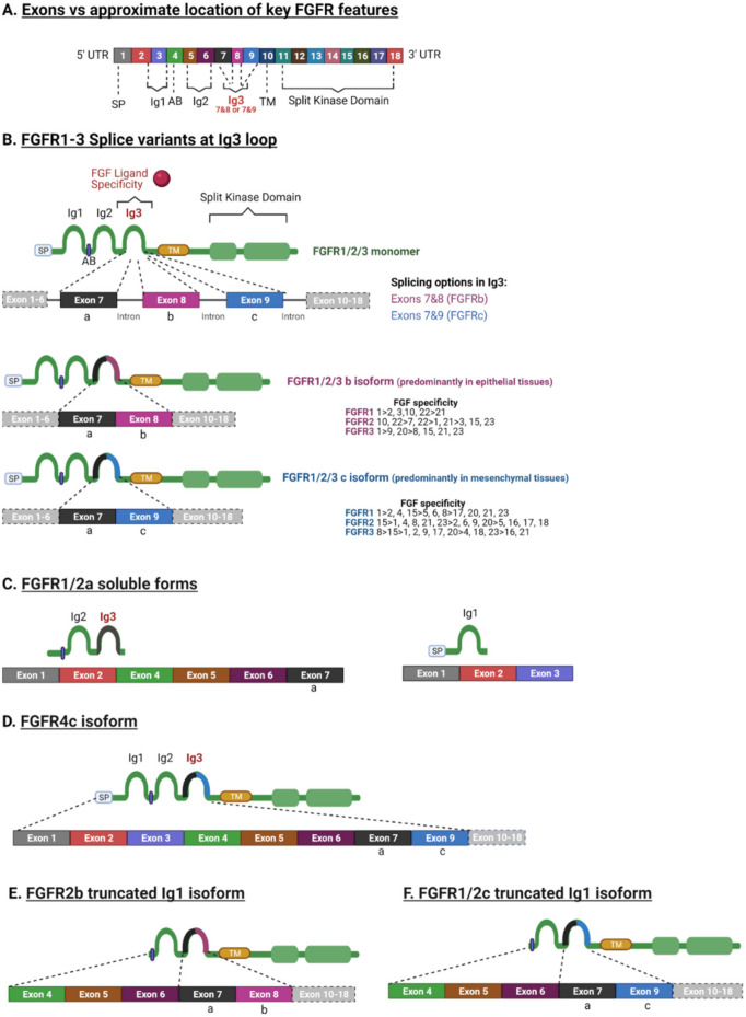
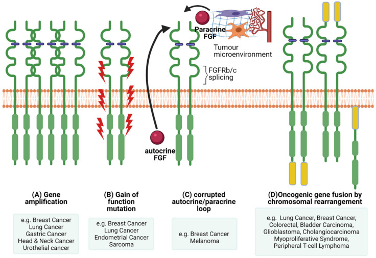
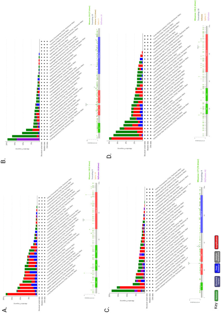
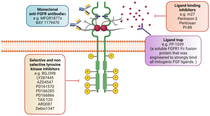
The pleiotropic effects of fibroblast growth factors (FGFs), the widespread expression of all seven signalling FGF receptors (FGFRs) throughout the body, and the dramatic phenotypes shown by many FGF/R knockout mice, highlight the diversity, complexity and functional importance of FGFR signalling. The FGF/R axis is critical during normal tissue development, homeostasis and repair. Therefore, it is not surprising that substantial evidence also pinpoints the involvement of aberrant FGFR signalling in disease, including tumourigenesis. FGFR aberrations in cancer include mutations, gene fusions, and amplifications as well as corrupted autocrine/paracrine loops. Indeed, many clinical trials on cancer are focusing on targeting the FGF/FGFR axis, using selective FGFR inhibitors, nonselective FGFR tyrosine kinase inhibitors, ligand traps, and monoclonal antibodies and some have already been approved for the treatment of cancer patients. The heterogeneous tumour microenvironment and complexity of FGFR signalling may be some of the factors responsible for the resistance or poor response to therapy with FGFR axis-directed therapeutic agents. In the present review we will focus on the structure and function of FGF(R)s, their common irregularities in cancer and the therapeutic value of targeting their function in cancer.

Keywords: FGFR inhibitors; FGFR mutations; FGFR signalling; cancer; fibroblast growth factor; targeting FGFR.


Conflict of interest statement

The authors declare no conflict of interest.

Figures

Figure 1
Figure 1
Fibroblast growth factor receptor (FGFR) structure and function. FGFR possesses three immunoglobulin-like domains (Ig1–3), an acid box (AB), a transmembrane domain (TM), and an intracellular tyrosine kinase domain that is split into two (TK1 and TK2). The FGF–FGFR complex is composed by two FGFs, two FGFRs and one heparin sulphate proteoglycan (HSPG). The TK domains are transphosphorylated upon ligand binding between Ig2-Ig3 and receptor dimerisation. This initiates the interaction between a network of downstream signalling molecules that can activate key pathways, such as MAPK, AKT, PLCγ, STAT1 and in turn regulate target genes involved in cell proliferation, migration, differentiation, survival, resistance to anticancer agents and neoangiogenesis. Signalling can be negatively regulated by SEF, FGFR-like 1 (FGFRL1), sprouty (SPRY) and MAPK phosphatase 1 and 3 (MKP1 and MKP3) at different levels. Created with BioRender.com (accessed on 26 September 2021).
Figure 2
Figure 2
Key FGFR splice variants and ligand specificity. (A) A generalised diagram representing the key FGFR features. (B) Splice variant at the Ig3 loop occurs in FGFR1–3. This splice variant is responsible for ligand binding specificity and is generated by alternative splicing at the Ig3. The first portion of the Ig3 is the exon “a” (exon 7) that is spliced to either exon “b” (exon 8) or exon “c” (exon 9) and then to the exon that encodes the TM domain. The “b” isoform is mainly expressed by epithelial tissues/cells, whereas the “c” isoform is expressed by mesenchymal tissues. FGFs have differential specificity to different isoforms. (C) Splice variants can generate soluble variants without TK activity, truncated to one or more Ig domains and missing the TM domain. Variants lacking the TK domain can heterodimerise with full length FGFRs to generate nonfunctional dimers and therefore act as down regulators [24]. (D) FGFR4 can generate a single isoform containing the “c” exon (exon 9) in the Ig3 domain. (E) FGFR2 can generate a splice variant missing Ig1 and Ig3 containing the “b” exon (exon 8). (F) FGFR1 and FGFR2 can also generate a splice variant with truncated Ig1 and Ig3 containing the “c” exon (exon 9). SP: signal peptide, Ig: Immunoglobulin, AB: acid box; TM: transmembrane domain, UTR: untranslated region. Created with BioRender.com (accessed on 26 September 2021).
Figure 3
Figure 3
FGFR dysregulations. There are several mechanisms of oncogenic FGFR function. (A) Gene amplification results in accumulation of FGFRs that usually translate into protein overexpression and activation of the FGFR axis. (B) Gain of function mutations can lead to constitutive FGFR activation with or without FGF binding. (C) Corrupted autocrine and paracrine loops, either via alternative splicing affecting ligand binding specificity or FGFRs getting overstimulated by FGFs produced in an autocrine fashion, by the cancer cells themselves, or by the tumour microenvironment, in a paracrine fashion. (D) Chromosomal rearrangements can lead to the creation of hybrid oncogenic FGFRs by fusing with binding partners at the carboxyl or amino termini. Created with BioRender.com (accessed on 7 November 2021).
Figure 4
Figure 4
FGFR1–4 genetic alteration in cancer. Genetic alterations by cancer type and mutations of FGFR1 (A), FGFR2 (B), FGFR3 (C) and FGFR4 (D) were found in 662 (~6%), 360 (~3%), 351 (~3%) and 285 (~3%) patients, respectively out of 10,953 in total using cBioPortal. Mutation diagram circles and histograms are coloured with respect to the corresponding mutation types: Green = gene mutations; purple = structural variants; blue = deep deletions; grey = multiple alterations; red = gene amplifications. The lollipop diagrams of each FGF receptor (FGFR1–4), below each histogram, represent the mutation types in relation to the gene location (i.e., missense, truncating, in-frame, splice, SV/fusion). In the case of different mutation types at a single position, the colour of the circle is determined with respect to the most frequent mutation type.
Figure 5
Figure 5
Targeting the FGFR axis. Aberrant FGFR signalling can contribute to cancer progression and therefore targeting FP-1039. can also block the FGF–FGFR interaction and therefore prohibit FGFR activation. In addition, ligand binding inhibitors that act as antagonists (e.g., PI-88 and sm27) can prevent FGFR activation. Monoclonal antibodies targeting specific FGFR isoforms (e.g., MFGR1877s by Genentech) can also have anti-tumour activity. Created with BioRender.com (accessed on 12 November 2021).

References

    1. Hanahan D., Weinberg R.A. Hallmarks of cancer: The next generation. Cell. 2011;144:646–674. doi: 10.1016/j.cell.2011.02.013. - DOI - PubMed
    1. Senga S.S., Grose R.P. Hallmarks of cancer-the new testament. Open Biol. 2021;11:200358. doi: 10.1098/rsob.200358. - DOI - PMC - PubMed
    1. Ornitz D.M. FGFs, heparan sulfate and FGFRs: Complex interactions essential for development. BioEssays. 2000;22:108–112. doi: 10.1002/(SICI)1521-1878(200002)22:2<108::AID-BIES2>3.0.CO;2-M. - DOI - PubMed
    1. Turner N., Grose R. Fibroblast growth factor signalling: From development to cancer. Nat. Rev. Cancer. 2010;10:116–129. doi: 10.1038/nrc2780. - DOI - PubMed
    1. Ornitz D.M., Xu J., Colvin J.S., McEwen D.G., MacArthur C.A., Coulier F., Gao G., Goldfarb M. Receptor specificity of the fibroblast growth factor family. J. Biol. Chem. 1996;271:15292–15297. doi: 10.1074/jbc.271.25.15292. - DOI - PubMed

LinkOut - more resources