Review
. 2021 Nov 15;13(22):5711.
doi: 10.3390/cancers13225711.

Fracture Risk Evaluation of Bone Metastases: A Burning Issue

Affiliations
Review

Fracture Risk Evaluation of Bone Metastases: A Burning Issue

Cyrille B Confavreux et al. Cancers (Basel). .

Abstract

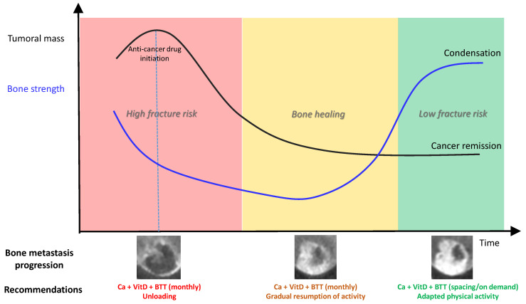
Major progress has been achieved to treat cancer patients and survival has improved considerably, even for stage-IV bone metastatic patients. Locomotive health has become a crucial issue for patient autonomy and quality of life. The centerpiece of the reflection lies in the fracture risk evaluation of bone metastasis to guide physician decision regarding physical activity, antiresorptive agent prescription, and local intervention by radiotherapy, surgery, and interventional radiology. A key mandatory step, since bone metastases may be asymptomatic and disseminated throughout the skeleton, is to identify the bone metastasis location by cartography, especially within weight-bearing bones. For every location, the fracture risk evaluation relies on qualitative approaches using imagery and scores such as Mirels and spinal instability neoplastic score (SINS). This approach, however, has important limitations and there is a need to develop new tools for bone metastatic and myeloma fracture risk evaluation. Personalized numerical simulation qCT-based imaging constitutes one of these emerging tools to assess bone tumoral strength and estimate the femoral and vertebral fracture risk. The next generation of numerical simulation and artificial intelligence will take into account multiple loadings to integrate movement and obtain conditions even closer to real-life, in order to guide patient rehabilitation and activity within a personalized-medicine approach.

Keywords: bone metastasis; finite element analysis; mirels’ score; neoplastic score; pathological fracture; spinal instability.


Conflict of interest statement

The authors declare no conflict of interest for this review. The funders had no role in the design and in the writing of the manuscript.

Figures

Figure 1
Figure 1
CT scan. Upper part: Multiplanar sagittal and axial reconstructions of the dorsal spine showing lytic lesions of myeloma involving T8 and T9 vertebrae. Lower part from left to right: progression of the size and condensation of a urothelial cancer bone metastasis under immunotherapy (avelumab), with initial lytic aspect and partial sclerosis of the lesion at 2 months. At 4 months, the sclerosis increases except in a focal region suspected of local progression. Confirmation of local progression with lysis of the sclerotic matrix at 6 months. FUP, follow-up.
Figure 2
Figure 2
Biomechanics. An example of an idealized mechanical test curve. A load-displacement curve is transformed into a stress–strain curve with geometric parameters. The green part is the linear part corresponding to the elastic domain. The orange part corresponds to the plastic deformation domain. The ultimate stress (in red) corresponds to the failure but can only be numerically determined using a failure criterion.
Figure 3
Figure 3
(A) Manual contour (in red) of a tumor, performed by a radiologist (annotator), (B) Patient-specific finite element model of proximal femur, with a specific representation of the metastasis.
Figure 4
Figure 4
(A) Low dose full-body scan in standing position obtained using the EOS system®. (B) Semi-automatic measurement of spinal statics and pelvic angles on front and profile images. (C) 3D-reconstruction of the whole vertebral column in standing position.
Figure 5
Figure 5
Conceptual model of bone strength progression according to osteolytic bone metastasis response to anti-cancer drug. BTT, bone-targeted treatments.

References

    1. Chiang A.C., Massague J. Molecular Basis of Metastasis. N. Engl. J. Med. 2008;359:2814–2823. doi: 10.1056/NEJMra0805239. - DOI - PMC - PubMed
    1. Thiery J. Epithelio-mesenchymal transformation and cancer. Nat. Rev. Cancer. 2002;2:442–454. doi: 10.1038/nrc822. - DOI - PubMed
    1. Kang Y., Siegel P.M., Shu W., Drobnjak M., Kakonen S.M., Cordón-Cardo C., Guise T.A., Massagué J. A multigenic program mediating breast cancer metastasis to bone. Cancer Cell. 2003;3:537–549. doi: 10.1016/S1535-6108(03)00132-6. - DOI - PubMed
    1. Clézardin P., Coleman R., Puppo M., Ottewell P., Bonnelye E., Paycha F., Confavreux C.B., Holen I. Bone metastasis: Mechanisms, therapies, and biomarkers. Physiol. Rev. 2021;101:797–855. doi: 10.1152/physrev.00012.2019. - DOI - PubMed
    1. Müller A., Homey B., Soto H., Ge N., Catron D., Buchanan M.E., McClanahan T., Murphy E.R., Yuan W., Wagner S.N., et al. Involvement of chemokine receptors in breast cancer metastasis. Nature. 2001;410:50–56. doi: 10.1038/35065016. - DOI - PubMed

LinkOut - more resources