Review
. 2021 Nov 15;13(22):5719.
doi: 10.3390/cancers13225719.

The Hepatic Sinusoid in Chronic Liver Disease: The Optimal Milieu for Cancer

Affiliations
Review

The Hepatic Sinusoid in Chronic Liver Disease: The Optimal Milieu for Cancer

Albert Gibert-Ramos et al. Cancers (Basel). .

Abstract

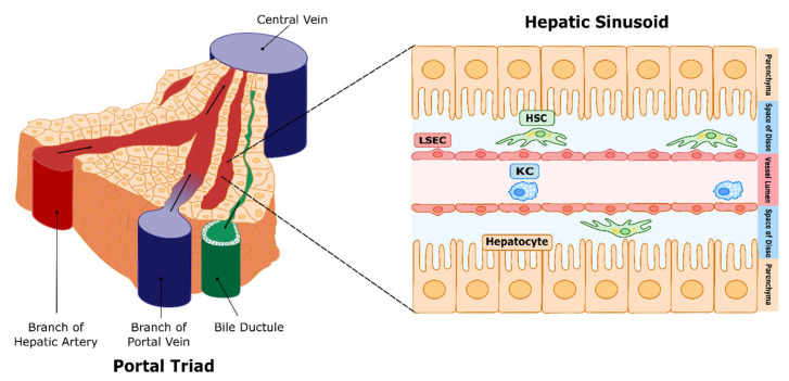
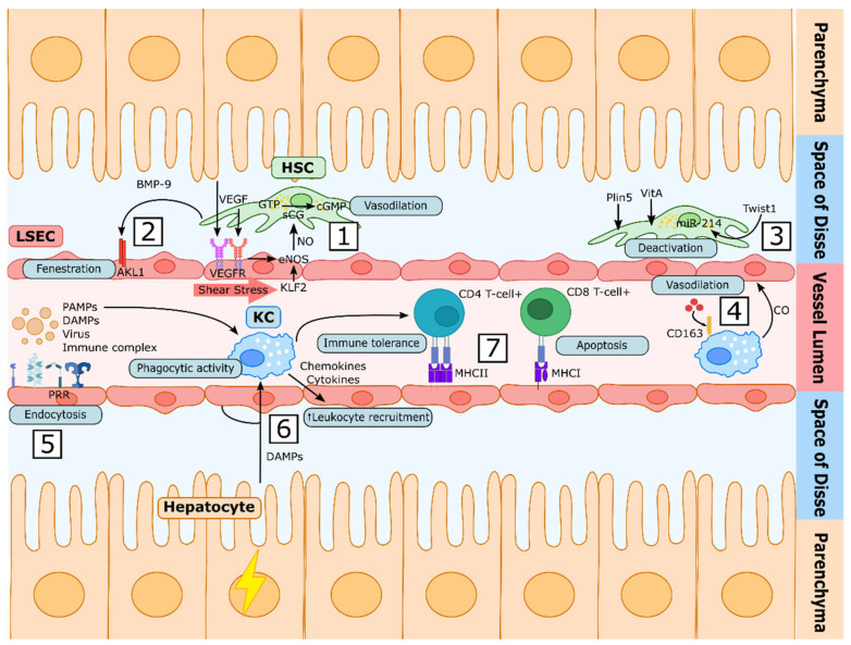
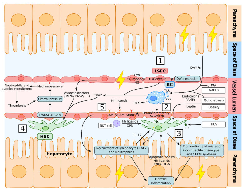
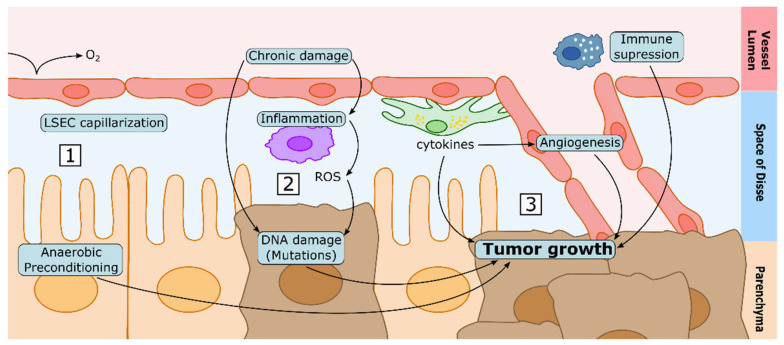
The liver sinusoids are a unique type of microvascular beds. The specialized phenotype of sinusoidal cells is essential for their communication, and for the function of all hepatic cell types, including hepatocytes. Liver sinusoidal endothelial cells (LSECs) conform the inner layer of the sinusoids, which is permeable due to the fenestrae across the cytoplasm; hepatic stellate cells (HSCs) surround LSECs, regulate the vascular tone, and synthetize the extracellular matrix, and Kupffer cells (KCs) are the liver-resident macrophages. Upon injury, the harmonic equilibrium in sinusoidal communication is disrupted, leading to phenotypic alterations that may affect the function of the whole liver if the damage persists. Understanding how the specialized sinusoidal cells work in coordination with each other in healthy livers and chronic liver disease is of the utmost importance for the discovery of new therapeutic targets and the design of novel pharmacological strategies. In this manuscript, we summarize the current knowledge on the role of sinusoidal cells and their communication both in health and chronic liver diseases, and their potential pharmacologic modulation. Finally, we discuss how alterations occurring during chronic injury may contribute to the development of hepatocellular carcinoma, which is usually developed in the background of chronic liver disease.

Keywords: CLD; HSC; Kupffer cell; LSEC; NASH; cirrhosis; hepatocellular carcinoma; portal hypertension.


Conflict of interest statement

The authors declare no conflict of interest.

Figures

Figure 1
Figure 1
Hepatic circulation and microcirculation. Representation of the liver circulation, with the portal triad, which includes the hepatic artery, which supplies oxygenated blood; the portal vein, carrying blood rich in nutrients from the small intestine; and the bile duct, which collects bile products secreted by hepatocytes. Blood then mixes along the sinusoids, which are the liver microvessels (right panel), and drains into the central vein, which leads to the vena cava. Liver sinusoidal endothelial cells (LSECs) constitute the walls of the microvessel. Hepatic stellate cells (HSCs) reside in the space defined between LSECs and hepatocytes (space of Disse), and act as the sinusoidal pericytes, while Kupffer cells (KCs) (resident macrophages) are located in the sinusoidal lumen.
Figure 2
Figure 2
Liver sinusoidal cells’ communication under physiological conditions. (1) In homeostatic conditions, LSECs sense VEGF through specific receptors (VEGFR). In parallel, mechanical shear stress induces Krüppel-like factor 2 (KLF2), altogether maintaining LSECs’ vasodilatory phenotype, inducing NO synthesis. NO activates the soluble guanylate cyclase (sGC) in HSCs, leading to vasodilation. (2) The endothelium is fenestrated in healthy conditions. Circulating bone morphogenetic protein (BMP-9) released by HSCs contributes to the maintenance of the fenestrae by its recognition through activin receptor-like kinase 1 (ALK1). (3) Perilipin 5 (Plin5) participates in the formation of vitamin A (VitA)-containing lipid droplets. In addition, quiescent HSCs secrete exosomes containing the transcription factor Twist1, which promotes HSCs quiescence autocrinally through the transcription of miRNA-214. (4) Kupffer cells (KCs) incorporate haemoglobin through the scavenger receptor CD163 and, by its degradation, produce vasoprotective products such as carbon monoxide (CO). (5) LSECs display high-affinity receptors which participate in innate immunity, such as pattern recognition receptors (PRRs), being capable of sensing pathogen-associated molecular patterns (PAMPs), damage-associated molecular patterns (DAMPs), viruses and other immune complexes. (6) KCs are responsible for the phagocytosis of bacteria or particle-associated antigens. Upon damage, KCs recognize PAMPs or DAMPS and produce cytokines and chemokines which increase the expression of adhesion molecules by LSECs, leading to leukocyte infiltration and activation. (7) LSECs can act as antigen-presenting cells, as they express major histocompatibility complexes I and II (MHC-I and MHC-II). Presentation to CD4+ T cells promotes their differentiation towards T regulatory (Treg) immunosuppressive cells, enhancing tolerance. On the other hand, LSEC antigen presentation to CD8+ T cells increases the programmed death of the CD8+ T cells suppressing the immune response.
Figure 3
Figure 3
Liver Sinusoid dysfunction during chronic liver disease. (1) During chronic liver injury, LSEC become dysfunctional, impairing the autophagy process, increasing the generation of reactive oxygen species (ROS), decreasing nitric oxide (NO) intrahepatic levels and synthetizing increased vasoconstrictors, which induces the activation of HSCs. Hepatic damage further induces LSECs defenestration through the degradation of caveolin-1. (2) Kupffer cells (KCs) are activated by damage-associated molecular patterns (DAMPs), pathogen-associated molecular patterns (PAMPs), free-fatty acids (FFA) and endotoxins via their toll-like receptors (TLR) and pattern recognition receptors (PRR). This induces the secretion of reactive oxygen species (ROS) and proinflammatory cytokines that, together with other proinflammatory molecules secreted by other cell types, activate HSCs (3) which will acquire a proliferative, migrating, procontractile and proinflammatory phenotype that will induce liver fibrosis and inflammation. This procontractile phenotype increases vascular tone (4), which further increases portal pressure, activating LSECs mechanosensors that induce the recruitment of neutrophiles and platelets, the accumulation of which produces thrombi that will further increase portal pressure. LSECs are also activated by hepatocyte-derived hedgehog (Hh) ligands and other proinflammatory mediators (5) secreted during the inflammatory and injury process, which—via adhesion molecules such as ICAM, VCAM and Stabilins—will recruit leukocytes to the liver tissue, further promoting fibrosis and inflammation.
Figure 4
Figure 4
Proposed mechanisms linking CLD and HCC development. (1) The capillarization of LSECs may lead to less oxygen diffusing to the space of Disse. Therefore, hepatocytes may be preconditioned to anaerobic metabolism in CLD, which may represent an advantage to tumoral cells in hypoxic conditions. (2) Chronic damage and the associated chronic inflammation are known causes of DNA damage, which may lead to cellular dedifferentiation and tumorigenesis. (3) Cytokines released by the different hepatic cell types in conditions of chronic liver damage may induce tumour growth directly, enhancing neovascularization, or by suppressing the immune system.

References

    1. McCuskey R.S.S. The hepatic microvascular system in health and its response to toxicants. Anat. Rec. 2008;291:661–671. doi: 10.1002/ar.20663. - DOI - PubMed
    1. Wisse E., de Zanger R.B., Charels K., van der Smissen P., McCuskey R.S. The liver sieve: Considerations concerning the structure and function of endothelial fenestrae, the sinusoidal wall and the space of disse. Hepatology. 1985;5:683–692. doi: 10.1002/hep.1840050427. - DOI - PubMed
    1. Parola M., Pinzani M. Liver fibrosis: Pathophysiology, pathogenetic targets and clinical issues. Mol. Aspects Med. 2019;65:37–55. doi: 10.1016/j.mam.2018.09.002. - DOI - PubMed
    1. Tsochatzis E.A., Bosch J., Burroughs A.K. Liver cirrhosis. Lancet. 2014;383:1749–1761. doi: 10.1016/S0140-6736(14)60121-5. - DOI - PubMed
    1. Forner A., Reig M., Bruix J. Hepatocellular carcinoma. Lancet. 2018;391:1301–1314. doi: 10.1016/S0140-6736(18)30010-2. - DOI - PubMed

LinkOut - more resources