. 2021 Nov 16;13(22):5739.
doi: 10.3390/cancers13225739.

Correlation of Soluble CD44 Expression in Saliva and CD44 Protein in Oral Leukoplakia Tissues

Affiliations

Correlation of Soluble CD44 Expression in Saliva and CD44 Protein in Oral Leukoplakia Tissues

Ingrīda Čēma et al. Cancers (Basel). .

Abstract

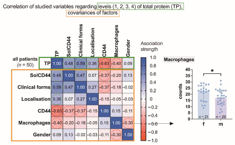
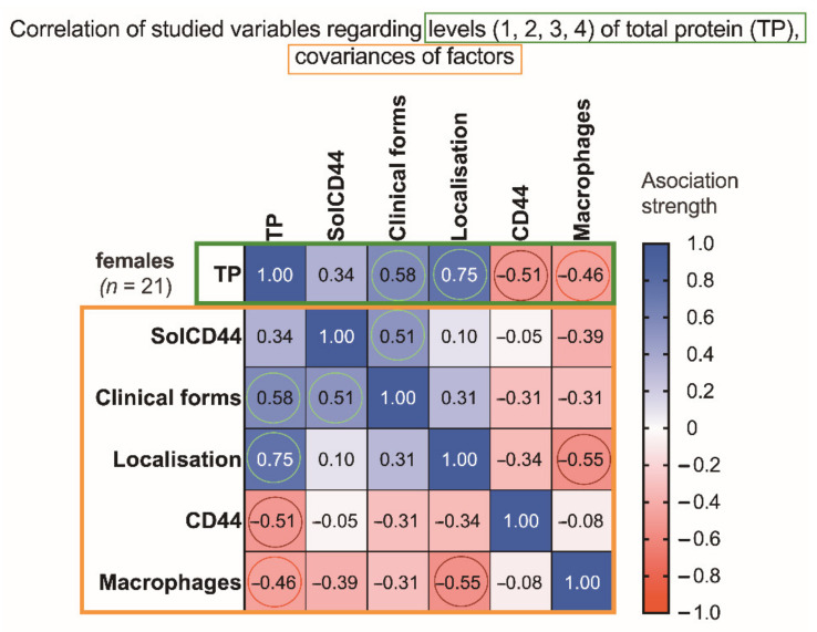
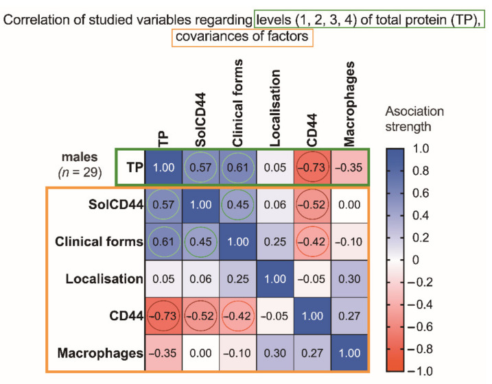
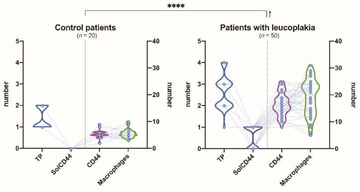
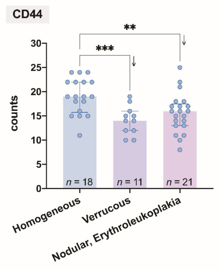
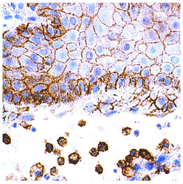
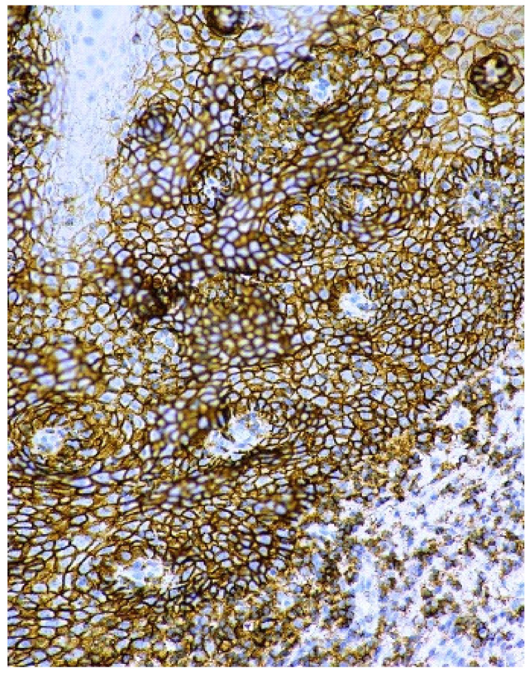
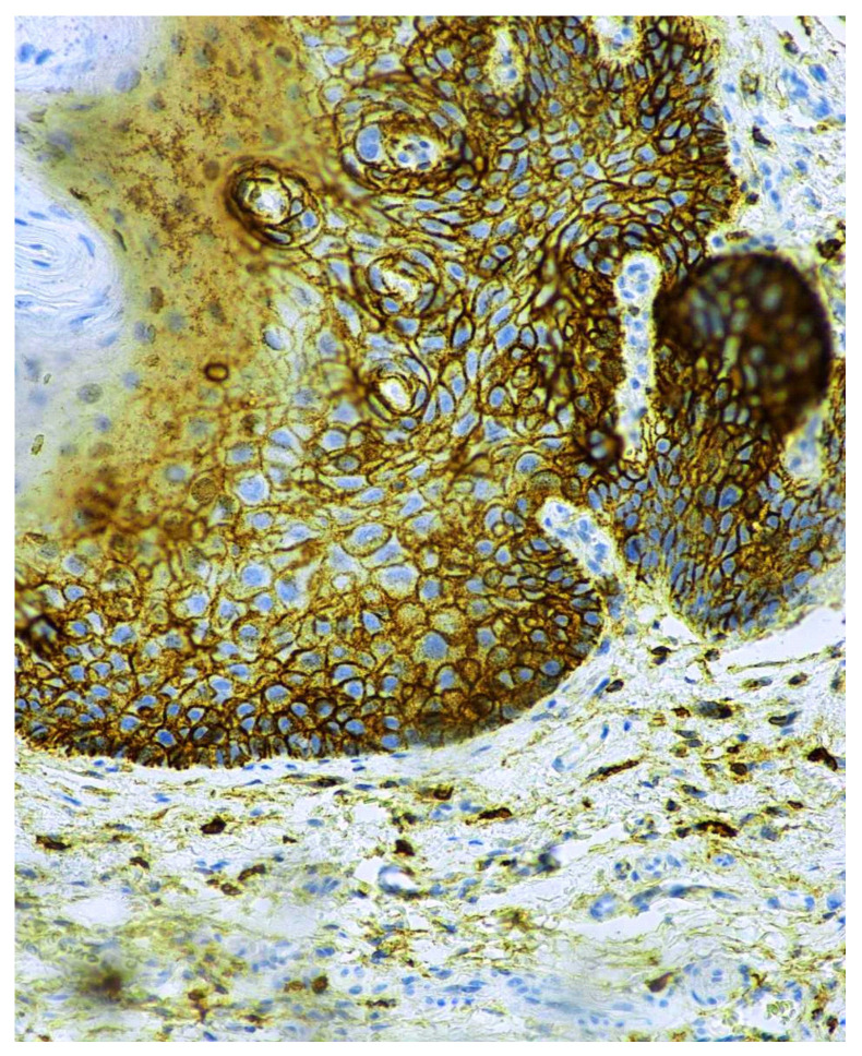
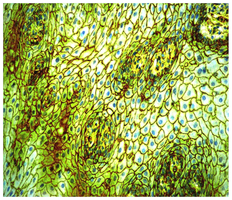
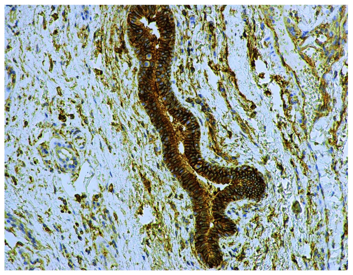
The aim of this study was to determine whether and how pan-CD44 protein expression in leukoplakia tissues correlates with positive SolCD44 test presence and their role in oral leukoplakia. SolCD44 and total protein expression in saliva were determined using an OncAlert® Oral Cancer Rapid test. Comparison of paired associations of total protein, SolCD44, mean number of CD44 expressed epithelial layers in leukoplakia tissue, and macrophages below the basement membrane between control group and patients with leukoplakia showed statistically significant results (p < 0.0001). It is shown that the total protein indicates low or elevated risk of possible malignant transformation processes in leukoplakia. Statistically significant differences between higher total protein level and clinical forms of oral leukoplakia (p < 0.0001), as well as CD44-labeled epithelial cell layer decrease (p < 0.0001), were found. This possibly points to the onset of the stemness loss in leukoplakia tissue. CD9 antigen expression in the exosomes of the oral epithelium explained the intercellular flow of SolCD44 and other fluids in the leukoplakia area. We conclude that the OncAlert® Oral Cancer Rapid test is a valuable screening method in daily clinical practice, in terms of complementing clinical diagnostics methods and to assess the potential for early malignancy.

Keywords: CD44 antigen; exosomes; oral leukoplakia; soluble CD44; total protein.


Conflict of interest statement

The authors declare no conflict of interest.

Figures

Figure 1
Figure 1
Association strength of studied variables: SolCD44, clinical forms of leukoplakia, localization of oral leukoplakia, mean number of CD44 expressed in epithelial layers in leukoplakia tissue, mononuclear cells (macrophages under the basal membrane), and gender of patients with leukoplakia (n = 50) regarding levels of total protein (and covariances). Spearman’s rank correlation matrix: numbers in squared cells show value of correlation coefficient showing association strength; colored circles denote the most significant associations (red, negative; green, positive). Negative association between the number of macrophages and gender (f, female; m, male) is additionally represented by the scatter plot on the right. * p < 0.05 (unpaired t-test). The arrow indicates a decrease in the average value of the corresponding group.
Figure 2
Figure 2
Association strength of studied variables: SolCD44, clinical form of leukoplakia, localization of oral leukoplakia, mean number of CD44 expressed in epithelial layers in leukoplakia tissue, mononuclear cells (macrophages under the basal membrane), and gender of patients with leukoplakia (n = 21) regarding levels of total protein (and covariances) in the female group. Spearman’s rank correlation matrix: numbers in squared cells show value of correlation coefficient, indicating association strength; colored circles denote most significant associations (red, negative; green, positive).
Figure 3
Figure 3
Association strength of studied variables: SolCD44, clinical form of leukoplakia, localization of oral leukoplakia, mean number of CD44 expressed in epithelial layers in leukoplakia tissue, mononuclear cells (macrophages under the basal membrane), and gender of patients with leukoplakia (n = 29) regarding levels of total protein (and covariances), in the male group. Spearman’s rank correlation matrix: numbers in squared cells show value of correlation coefficient, indicating association strength; colored circles denote most the significant associations (red, negative; green, positive).
Figure 4
Figure 4
Comparison of paired associations of total protein, SolCD44, mean number of CD44 expressed in epithelial layers in leukoplakia tissue, and macrophages in lamina propria between control group and patients with leukoplakia. **** p < 0.0001 (repeated-measures two-way ANOVA with two-stage linear step-up procedure of Benjamini, Krieger, and Yekutieli as post hoc procedure) indicates the level of statistical significance of between-group difference, regarding the analyzed parameters. The arrow indicates an increase in the average values of the corresponding group compared to the control.
Figure 5
Figure 5
Comparison of the mean number of CD44 expressed in epithelial layers in leukoplakia tissue and clinical forms of oral leukoplakia. Levels of statistical significance: ** p < 0.01, *** p < 0.001 (Ordinary one-way ANOVA with two-stage linear step-up procedure of Benjamini, Krieger, and Yekutieli as a post hoc procedure). The arrows indicate a decrease in the average value of the corresponding groups.
Figure 6
Figure 6
Immunohistochemical visualization of the CD44 marker in epithelium and mononuclear cells of lamina propria of healthy oral mucosa. Immunoperoxidase, anti-CD44, original magnification 400×.
Figure 7
Figure 7
Overexpression of CD44 protein in the cell membranes of epithelium in oral leukoplakia. Immunoperoxidase, anti-CD44, original magnification 200×.
Figure 8
Figure 8
Membranous and intra-cytoplasmatic expression of the CD44 antigen in non-homogenous oral leukoplakia. Immunoperoxidase, anti-CD44, original magnification 200×.
Figure 9
Figure 9
Immunohistochemical visualization of the CD9 marker in the cell membranes of epithelium and its exosomes of oral leukoplakia. Immunoperoxidase, anti- CD9, original magnification 400×.
Figure 10
Figure 10
CD9 antigen expression in the lumen microparticiples and the ductal epithelium of small salivary glands under oral leukoplakia. Immunoperoxidase, anti-CD9, original magnification 200×.

Similar articles

Cited by

References

    1. Sung H., Ferlay J., Siegel R.L., Laversanne M., Soerjomataram I., Jemal A., Bray F. Global Cancer Statistics 2020: GLOBOCAN Estimates of Incidence and Mortality Worldwide for 36 Cancers in 185 Countries. CA Cancer J. Clin. 2021;71:209–249. doi: 10.3322/caac.21660. - DOI - PubMed
    1. Diz P., Meleti M., Diniz-Freitas M., Vescovi P., Warnakulasuriya S., Johnson N.W., Kerr A.R. Oral and pharyngeal cancer in Europe: Incidence, mortality and trends as presented to the Global Oral Cancer Forum. Transl. Res. Oral Oncol. 2017;2:2057178X17701517. doi: 10.1177/2057178X17701517. - DOI
    1. Roza A.L.O.C., Kowalski L.P., William W.N., de Castro G., Chaves A.L.F., Araújo A.L.D., Ribeiro A.C.P., Brandão T.B., Lopes M.A., Vargas P.A., et al. Oral leukoplakia and erythroplakia in young patients: A systematic review. Oral Surg. Oral Med. Oral Pathol. Oral Radiol. 2021;131:73–84. doi: 10.1016/j.oooo.2020.09.002. - DOI - PubMed
    1. Petti S. Pooled estimate of world leukoplakia prevalence: A systematic review. Oral Oncol. 2003;39:770–780. doi: 10.1016/S1368-8375(03)00102-7. - DOI - PubMed
    1. Mello F.W., Miguel A.F.P., Dutra K.L., Porporatti A.L., Warnakulasuriya S., Guerra E.N.S., Rivero E.R.C. Prevalence of oral potentially malignant disorders: A systematic review and meta-analysis. J. Oral Pathol. Med. 2018;47:633–640. doi: 10.1111/jop.12726. - DOI - PubMed

LinkOut - more resources