. 2021 Nov 18;13(22):5784.
doi: 10.3390/cancers13225784.

On Efficacy of Microwave Ablation in the Thermal Treatment of an Early-Stage Hepatocellular Carcinoma

Affiliations

On Efficacy of Microwave Ablation in the Thermal Treatment of an Early-Stage Hepatocellular Carcinoma

Branislav Radjenović et al. Cancers (Basel). .

Abstract

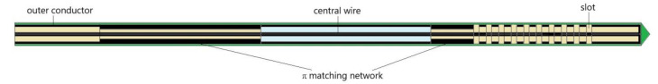
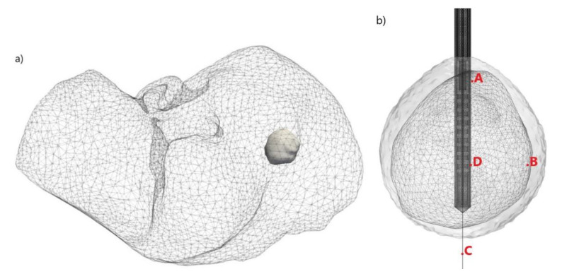
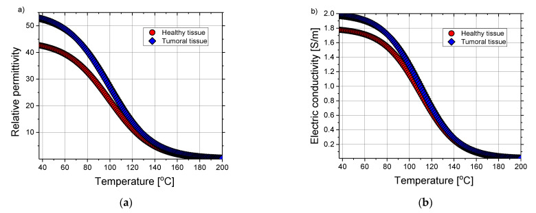
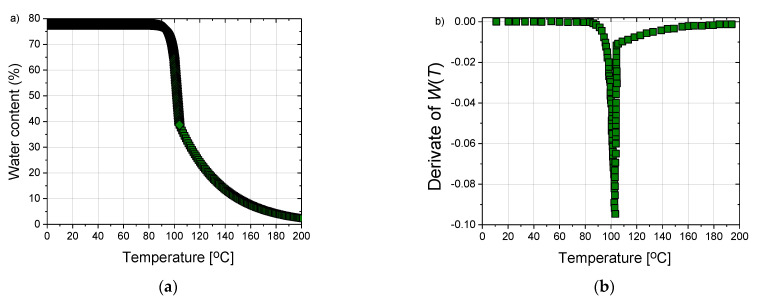
Microwave ablation at 2.45 GHz is gaining popularity as an alternative therapy to hepatic resection with a higher overall survival rate than external beam radiation therapy and proton beam therapy. It also offers better long-term recurrence-free overall survival when compared with radiofrequency ablation. To improve the design and optimization of microwave ablation procedures, numerical models can provide crucial information. A three-dimensional model of the antenna and targeted tissue without homogeneity assumptions are the most realistic representation of the physical problem. Due to complexity and computational resources consumption, most of the existing numerical studies are based on using two-dimensional axisymmetric models to emulate actual three-dimensional cancers and surrounding tissue, which is often far from reality. The main goal of this study is to develop a fully three-dimensional model of a multislot microwave antenna immersed into liver tissue affected by early-stage hepatocellular carcinoma. The geometry of the tumor is taken from the 3D-IRCADb-01 liver tumors database. Simulations were performed involving the temperature dependence of the blood perfusion, dielectric and thermal properties of both healthy and tumoral liver tissues. The water content changes during the ablation process are also included. The optimal values of the input power and the ablation time are determined to ensure complete treatment of the tumor with minimal damage to the healthy tissue. It was found that a multislot antenna is designed to create predictable, large, spherical zones of the ablation that are not influenced by varying tissue environments. The obtained results may be useful for determining optimal conditions necessary for microwave ablation to be as effective as possible for treating early-stage hepatocellular carcinoma, with minimized invasiveness and collateral damages.

Keywords: hepatocellular carcinoma; microwave ablation; necrotic tissue.


Conflict of interest statement

The authors declare no conflict of interest.

Figures

Figure 1
Figure 1
Schematic view of the 10-slot microwave antenna with an impedance match network. Black, green, light blue, and brown colors correspond to conducting material, Teflon, air, and dielectric, respectively. The width of the slot is 0.6 mm, while the spacing between slots is 0.8 mm.
Figure 2
Figure 2
Three-dimensional (a) simulation model of the liver (triangulated surface) and an early-stage HCC (solid surface) corresponding to patient 16 in the 3D-IRCADb-01 database [70] and (b) configuration of the antenna inserted into biological tissue and positions of four test points (A, B, C, D).
Figure 3
Figure 3
The temperature dependence of (a) relative permittivity and (b) electric conductivity of the healthy (red circles) and tumoral liver tissue (blue diamonds). Plots are obtained by using expressions (2) and (3) with coefficients taken from the literature [68].
Figure 4
Figure 4
(a) The time dependence of the water content W(T) of the tissue according to expression (8), taken from reference [85] and (b) the first derivate of W(T) which is then used to calculate an effective specific heat given by Equation (9).
Figure 5
Figure 5
Results of (a) two-dimensional axial-symmetric and (b) full three-dimensional simulation models of the temperature (expressed in °C) distributions in the liver tissue after 600 s of microwave ablation at a frequency 2.45 GHz and the input power of 10 W.
Figure 6
Figure 6
Isocontours represent the totally ablated regions (gray surfaces) around the liver tumor [70] (triangulated surface) exposed to 600 s of microwave ablation at 2.45 GHz and the three values of the input power (10 W, 13 W, and 15 W).
Figure 7
Figure 7
(a) x–y, (b) y–z, and (c) x–z cut planes of the total power dissipation density (expressed in W/m3) calculated for the liver tumor [70] (plotted as triangulated surface) exposed to microwave frequency of 2.45 GHz and input power of 13 W after 600 s.
Figure 8
Figure 8
Temporal evolution of the temperature (in °C) when an early-stage HCC [70] is treated by microwave ablation at a frequency of 2.45 GHz and input power of 13 W in y–z cut plane. The boundary of the tumor tissue is marked by the black line.
Figure 9
Figure 9
(a) Three-dimensional plot of the liver tumor [70] represented by triangulated surface and isocontours that correspond to the temperature of 40 °C (light gray), 60 °C (light brown), and 70 °C (brown). (b) The time dependence of the temperature calculated at test points (A, B, C, and D) is marked in Figure 4b.
Figure 10
Figure 10
The time evolution of the fraction of necrotic tissue exposed to the microwave ablation at a frequency of 2.45 GHz and the input power of 13 W. The boundary of the tumoral tissue is marked by the black line.
Figure 11
Figure 11
(a) The liver tumor [70] is shown as triangulated surface and isocontours corresponding to the fractions of damage of 0.3 (violet), 0.5 (blue), 0.75 (dark green), and 1 (dark gray). (b) The time evolution of the necrotic tissue was calculated at four test points (A, B, C, and D) marked in Figure 4b.

References

    1. Ananthakrishnan A., Gogineni V., Kia Saeian K. Epidemiology of Primary and Secondary Liver Cancers. Semin. Intervent. Radiol. 2006;23:47–63. doi: 10.1055/s-2006-939841. - DOI - PMC - PubMed
    1. Vonlaufen A., Phillips P.A., Xu Z., Goldstein D., Pirola R.C., Wilson J.S., Apte M.V. Pancreatic stellate cells and pancreatic cancer cells: An unholy alliance. Cancer Res. 2008;68:7707–7710. doi: 10.1158/0008-5472.CAN-08-1132. - DOI - PubMed
    1. Yamashita T., Wang X.W. Cancer stem cells in the development of liver cancer. J. Clin. Investig. 2013;123:1911–1918. doi: 10.1172/JCI66024. - DOI - PMC - PubMed
    1. Liu H., Zhang W., Jia Y., Yu Q., Grau G.E., Peng L., Ran Y., Yang Z., Deng H., Lou J. Single-cell clones of liver cancer stem cells have the potential of differentiating into different types of tumor cells. Cell Death Dis. 2013;4:e857. doi: 10.1038/cddis.2013.340. - DOI - PMC - PubMed
    1. Willacy H. Primary Liver Cancer. 2018. [(accessed on 8 August 2021)]. Available online: https://patient.info/cancer/primary-liver-cancer-leaflet.

LinkOut - more resources