. 2021 Nov 19;13(22):5807.
doi: 10.3390/cancers13225807.

Dual-Wavelength Fluorescence Monitoring of Photodynamic Therapy: From Analytical Models to Clinical Studies

Affiliations

Dual-Wavelength Fluorescence Monitoring of Photodynamic Therapy: From Analytical Models to Clinical Studies

Mikhail Kirillin et al. Cancers (Basel). .

Abstract

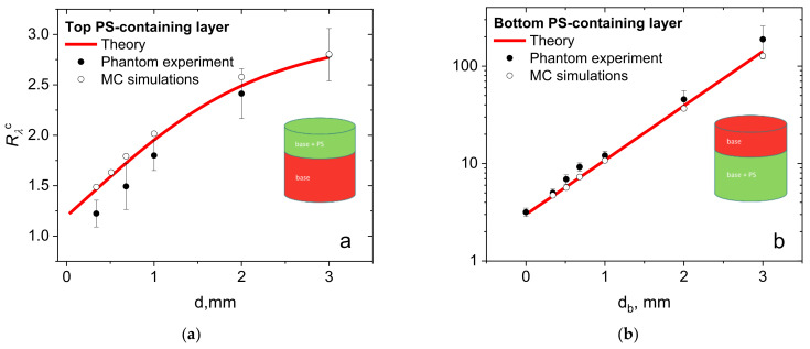
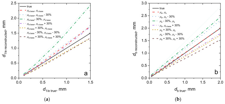
Fluorescence imaging modalities are currently a routine tool for the assessment of marker distribution within biological tissues, including monitoring of fluorescent photosensitizers (PSs) in photodynamic therapy (PDT). Conventional fluorescence imaging techniques provide en-face two-dimensional images, while depth-resolved techniques require complicated tomographic modalities. In this paper, we report on a cost-effective approach for the estimation of fluorophore localization depth based on dual-wavelength probing. Owing to significant difference in optical properties of superficial biotissues for red and blue ranges of optical spectra, simultaneous detection of fluorescence excited at different wavelengths provides complementary information from different measurement volumes. Here, we report analytical and numerical models of the dual-wavelength fluorescence imaging of PS-containing biotissues considering topical and intravenous PS administration, and demonstrate the feasibility of this approach for evaluation of the PS localization depth based on the fluorescence signal ratio. The results of analytical and numerical simulations, as well as phantom experiments, were translated to the in vivo imaging to interpret experimental observations in animal experiments, human volunteers, and clinical studies. The proposed approach allowed us to estimate typical accumulation depths of PS localization which are consistent with the morphologically expected values for both topical PS administration and intravenous injection.

Keywords: Monte Carlo simulations; animal studies; chlorin-based photosensitizers; clinical studies; dual-wavelength fluorescence imaging; light transport; optical phantoms; photodynamic therapy.


Conflict of interest statement

The authors declare no competing interests.

Figures

Figure 1
Figure 1
Dual-wavelength fluorescence imaging with chlorin e6 based photosensitizers. (a) Absorption and fluorescence spectra of chlorin e6-based PS and typical spectra of scattering and absorption coefficients of human dermis (*adopted from Salomatina et al. [23]); arrows show fluorescence excitation wavelengths employed in the proposed dual-wavelength imaging; (b) principles of FI with dual-wavelength excitation for topical PS administration (left) and intravenous PS injection (right).
Figure 2
Figure 2
Schematics of the considered cases of PS distribution in the tissue mimicking basic examples of topical or systemic PS administration: (a) PS is distributed uniformly in the upper layer of biotissue of thickness d; (b) PS is distributed uniformly within the semispace below the biotissue layer of thickness db; (c) PS concentration exponentially decreases in depth with the 1/e decay depth d1/e; and (d) a PS layer with an exponentially decaying concentration with the scale d1/e is covered by the biotissue layer of thickness db. The dependence CPS(z) illustrates the PS concentration in-depth profile.
Figure 3
Figure 3
Reconstructed spectra of the reduced scattering (a) and absorption (b) coefficients of the base tissue phantom and the phantom with PS, and the absorption spectrum of the water-based photosensitizer gel Revixan Derma dissolved in purified water (c). Dashed lines show the fluorescence excitation (405 nm, 660 nm) and emission detection (760 nm) wavelengths.
Figure 4
Figure 4
Analytical (theory) and numerically simulated (MC) dependencies of the fluorescence signals at different excitation wavelengths (ac) and signal ratios Rλ (df) on the characteristic PS localization depth within human dermis mimicking biotissue, which model different methods of PS administration: (a,d) topical PS administration—uniform PS distribution within the top layer; (b,e) systemic PS injection—uniform PS distribution in the bottom layer; (c,f) topical PS administration—exponential PS concentration in-depth profile.
Figure 5
Figure 5
Experimentally measured fluorescence signal ratio for the agarose biotissue phantoms with (a) a PS-containing top layer and (b) a PS-containing bottom layer versus the PS localization depth, corresponding analytical dependencies and results of the Monte Carlo simulations.
Figure 6
Figure 6
Reconstructed values of the fluorophore localization depth versus the true localization depth, depending on the medium optical properties employed for the reconstruction for cases of topical PS administration (a) and intravenous injection (b).
Figure 7
Figure 7
Normalized fluorescence signal ratio values of Rλc detected after the PS accumulation (a) and the corresponding PS localization depths (b) for topical application (TA) and intravenous injection (SA) in laboratory animals (CT26 tumor model in Balb/c mice, inner surface of the rabbit ear), human volunteers (normal human skin) and in patients (actinic keratosis, BCC). In the brackets, total number of time points, treatment sites and independent species in the group is shown.
Figure 8
Figure 8
Normalized fluorescence signal ratios prior to and after PDT procedures obtained in animal studies (inner surface of rabbit ear in norm and CT26 tumor model in Balb/c mice) for topical application (a) and intravenous injection (b). The PDT regimes’ abbreviations below the bars indicate the therapeutic wavelength (‘r’ = 660 nm, ‘b’ = 405 nm, ‘rb’ = 660 nm + 405 nm) and dose in J/cm2.
Figure 9
Figure 9
Typical fluorescence images of the BCC node site prior to (a,c) and after (b,d) the PDT procedure delivered at λ = 660 nm, with a dose of 150 J/cm2 acquired at probing wavelengths of 405 (a,b) and 660 (c,d) nm.
Figure 10
Figure 10
Fluorescence signal ratio prior to and after the PDT procedures obtained in the treatment of actinic keratosis (AK) with topical PS administration and basal cell carcinoma (BCC) with intravenous PS injection. The PDT regimes’ abbreviations below the bars show the therapeutic wavelength (‘r’ = 660 nm, ‘b’ = 405 nm, ‘rb’ = 660 nm + 405 nm) and dose in Joules.
Figure 11
Figure 11
PS localization depth changes, Δdb measured in laboratory animals and in patients as a result of the PDT procedure (TA—topical application; SA—systemic administration). In the brackets, total number of procedures, treatment sites and independent species in the group is shown.
Figure 12
Figure 12
Normalized fluorescence signal ratio, Rλc, summarized over all of the measurements in laboratory animals, human volunteers and patients upon PS administration or/and in the course of a PDT procedure (TA—topical application; SA—systemic administration). In the brackets, total number of time points, treatment sites and independent species in the group is shown.

Similar articles

Cited by

References

    1. Andersson-Engels S., af Klinteberg C., Svanberg K., Svanberg S. In vivo fluorescence imaging for tissue diagnostics. Phys. Med. Biol. 1997;42:815. doi: 10.1088/0031-9155/42/5/006. - DOI - PubMed
    1. Frangioni J.V. In vivo near-infrared fluorescence imaging. Curr. Opin. Chem. Biol. 2003;7:626–634. doi: 10.1016/j.cbpa.2003.08.007. - DOI - PubMed
    1. Bao X., Yuan Y., Chen J., Zhang B., Li D., Zhou D., Jing P., Xu G., Wang Y., Holá K. In vivo theranostics with near-infrared-emitting carbon dots—highly efficient photothermal therapy based on passive targeting after intravenous administration. Light Sci. Appl. 2018;7:1–11. doi: 10.1038/s41377-018-0090-1. - DOI - PMC - PubMed
    1. He S., Song J., Qu J., Cheng Z. Crucial breakthrough of second near-infrared biological window fluorophores: Design and synthesis toward multimodal imaging and theranostics. Chem. Soc. Rev. 2018;47:4258–4278. doi: 10.1039/C8CS00234G. - DOI - PubMed
    1. Celli J.P., Spring B.Q., Rizvi I., Evans C.L., Samkoe K.S., Verma S., Pogue B.W., Hasan T. Imaging and photodynamic therapy: Mechanisms, monitoring, and optimization. Chem. Rev. 2010;110:2795–2838. doi: 10.1021/cr900300p. - DOI - PMC - PubMed