. 2021 Oct 20;10(11):2805.
doi: 10.3390/cells10112805.

Gfi1 Loss Protects against Two Models of Induced Diabetes

Affiliations

Gfi1 Loss Protects against Two Models of Induced Diabetes

Tiziana Napolitano et al. Cells. .

Abstract

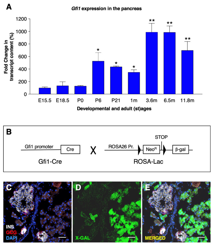
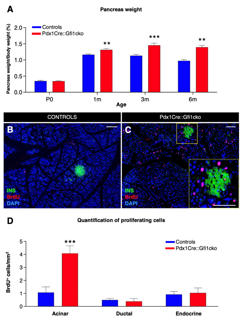
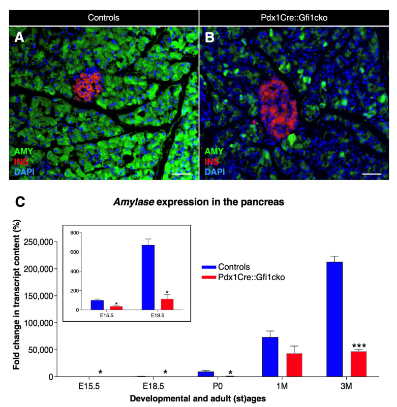
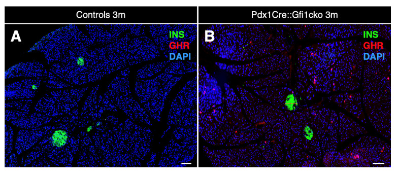
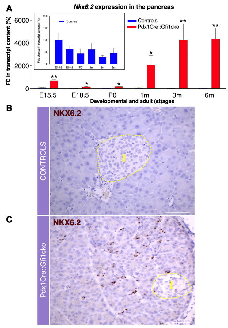
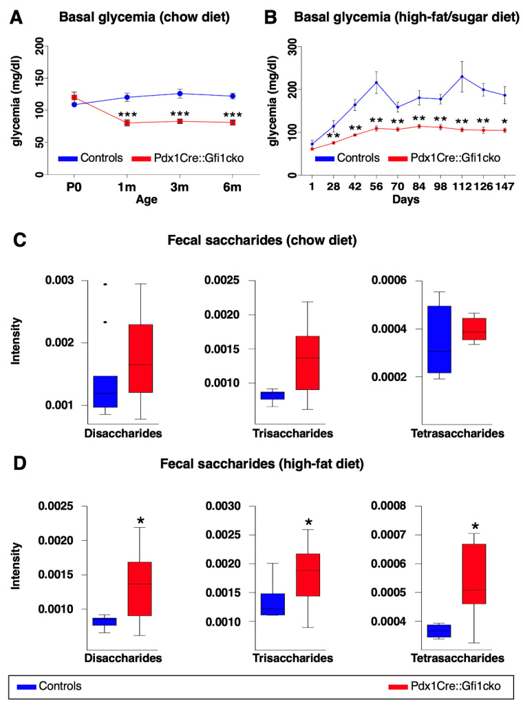
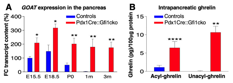
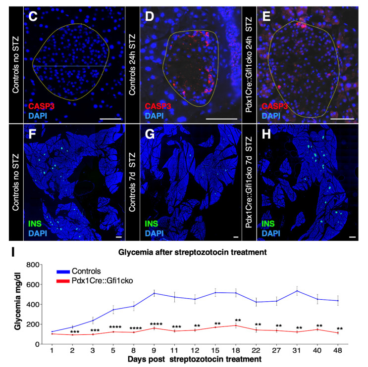
Background: Although several approaches have revealed much about individual factors that regulate pancreatic development, we have yet to fully understand their complicated interplay during pancreas morphogenesis. Gfi1 is transcription factor specifically expressed in pancreatic acinar cells, whose role in pancreas cells fate identity and specification is still elusive. Methods: In order to gain further insight into the function of this factor in the pancreas, we generated animals deficient for Gfi1 specifically in the pancreas. Gfi1 conditional knockout animals were phenotypically characterized by immunohistochemistry, RT-qPCR, and RNA scope. To assess the role of Gfi1 in the pathogenesis of diabetes, we challenged Gfi1-deficient mice with two models of induced hyperglycemia: long-term high-fat/high-sugar feeding and streptozotocin injections. Results: Interestingly, mutant mice did not show any obvious deleterious phenotype. However, in depth analyses demonstrated a significant decrease in pancreatic amylase expression, leading to a diminution in intestinal carbohydrates processing and thus glucose absorption. In fact, Gfi1-deficient mice were found resistant to diet-induced hyperglycemia, appearing normoglycemic even after long-term high-fat/high-sugar diet. Another feature observed in mutant acinar cells was the misexpression of ghrelin, a hormone previously suggested to exhibit anti-apoptotic effects on β-cells in vitro. Impressively, Gfi1 mutant mice were found to be resistant to the cytotoxic and diabetogenic effects of high-dose streptozotocin administrations, displaying a negligible loss of β-cells and an imperturbable normoglycemia. Conclusions: Together, these results demonstrate that Gfi1 could turn to be extremely valuable for the development of new therapies and could thus open new research avenues in the context of diabetes research.

Keywords: amylase; ghrelin; high-fat diet; mouse model; pancreas development.


Conflict of interest statement

The authors declare no conflict of interest.

Figures

Figure 1
Figure 1
Gfi1 expression in the mouse pancreas. The expression levels of Gfi1 were assessed by RT-qPCR using RNAs from whole pancreata from control mice (n = 6) at different developmental and adult (st) ages (* p < 0.05, ** p < 0.01). (A). Antibodies specifically recognizing Gfi1 are not commercially available. The Gfi1 expression pattern was therefore determined using a transgenic mouse line expressing a reporter gene under the control of Gfi1 promoter (B). Specifically, Gfi1-Cre animals (the Cre recombinase being knocked-in into the Gfi1 locus) were crossed with the Rosa26-β-gal mice (harboring a transgene composed of the ubiquitous Rosa26 promoter upstream of a LoxP-STOP-LoxP cassette followed by the β-galactosidase cDNA). An X-gal staining was performed on 4-month-old Gfi1Cre::RosaLac animals (the blue channel was converted in green to increase the image readability) to reveal β-galactosidase activity. Objective magnification: 20×. Scale bar: 50 μm (CE).
Figure 2
Figure 2
Loss of Gfi1 is associated with a dramatic increase in proliferation rate within the acinar compartment. Measurement of pancreas weight normalized to animal body weight at birth and 3 adult ages. (n = 8 animals per genotype per (st) age) (** p < 0.01, *** p < 0.001) (A). Adult Pdx1Cre::Gfi1cko and controls were provided with BrdU for 5 days. Subsequently, pancreatic sections were analyzed by immunofluorescence. Objective magnification: 20×. Scale bar: 100 μm (B,C). Quantitative analyses of BrdU-labelled cells in different pancreatic compartments (n = 4 animals per genotype) (*** p < 0.001) (D).
Figure 3
Figure 3
Important decrease in amylase expression in Pdx1Cre::Gfi1cko animals. Immunohistochemical analyses of Pdx1Cre::Gfi1cko and control mice using antibodies recognizing insulin (red) amylase (green) and DAPI (blue). Objective magnification: 20×. Scale bar: 50 μm (A,B). Amylase expression levels were assessed by RT-qPCR at different developmental and adult ages. Insert graph shows amylase expression at E15.5 and E18.5 of both Gfi1-conditional knockouts and controls. (n = 5 animals per genotype per (st) age) (* p < 0.05, *** p < 0.001) (C).
Figure 4
Figure 4
Ghrelin is misexpressed in Pdx1Cre::Gfi1cko adult pancreas. Immunohistochemical assessment of ghrelin-labeled cells in 3-month-old Pdx1Cre::Gfi1cko adult acinar cells. Objective magnification: 10×. Scale bar: 100 μm (A,B). Immunohistochemical analyses of amylase- and ghrelin-expressing cells in Gfi1-deficient pancreatic tissue. Objective magnification: 20×. Scale bar: 20 μm (C). Quantification of amylase and ghrelin co-immunoexpressing cells in Gfi1-deficient pancreatic acinar compartment (n = 253 ghrelin+ cells) (E). Phenotypical evaluations of ghrelin and BrdU labelled cells by immunofluorescence in Pdx1Cre::Gficko adult pancreas. Objective magnification: 20×. Scale bar: 50 μm (D). Quantitative evaluation of pancreatic ghrelin expression by RT-qPCR at different developmental and adult (st)ages (n = 5 animals per genotype per (st) age) (*** p < 0.001) (F).
Figure 4
Figure 4
Ghrelin is misexpressed in Pdx1Cre::Gfi1cko adult pancreas. Immunohistochemical assessment of ghrelin-labeled cells in 3-month-old Pdx1Cre::Gfi1cko adult acinar cells. Objective magnification: 10×. Scale bar: 100 μm (A,B). Immunohistochemical analyses of amylase- and ghrelin-expressing cells in Gfi1-deficient pancreatic tissue. Objective magnification: 20×. Scale bar: 20 μm (C). Quantification of amylase and ghrelin co-immunoexpressing cells in Gfi1-deficient pancreatic acinar compartment (n = 253 ghrelin+ cells) (E). Phenotypical evaluations of ghrelin and BrdU labelled cells by immunofluorescence in Pdx1Cre::Gficko adult pancreas. Objective magnification: 20×. Scale bar: 50 μm (D). Quantitative evaluation of pancreatic ghrelin expression by RT-qPCR at different developmental and adult (st)ages (n = 5 animals per genotype per (st) age) (*** p < 0.001) (F).
Figure 5
Figure 5
Gfi1 is crucially involved in the regulation of Nkx6.2 expression in mouse pancreas. Expression levels of Nkx6.2 measured by RT-qPCR during the embryonic development and adulthood in the pancreas of Pdx1Cre::Gfi1cko animals and age-matched controls (n = 5 animals per genotype per (st) age). Insert graph shows Nkx6.2 expression exclusively in control pancreatic samples. (* p < 0.05, ** p < 0.01) (A). Nkx6.2 expression pattern was assessed by RNA scope in adult Gfi1 loss of function and control pancreatic sections. Yellow dash line indicates the islet compartment. Objective magnification: 25×. Scale bar: 50 μm (B,C).
Figure 6
Figure 6
Pdx1Cre::Gfi1cko mice are resistant to high-fat/high-sugar diet-induced hyperglycemia. Basal glycemia was assessed in control and Gfi1-deficient mice (n = 8) at different ages. (*** p < 0.001) (A). Control and Gfi1 mutant mice were fed with HFCD (n = 7 animals per genotype) and their glycemia was monitored for 5 months. (* p < 0.05, ** p < 0.01) (B). Semiquantitative assessment of fecal saccharides by mass spectrometry was performed using samples of Pdx1Cre::Gfi1cko mice and controls fed with chow diet (C) and HFCD (D). (chow diet, n = 11 for control and 16 for Pdx1Cre::Gfi1cko samples; HFCG, n = 5 for control and 12 for Pdx1Cre::Gfi1cko samples) (* p < 0.05).
Figure 7
Figure 7
Pdx1Cre::Gfi1cko mice are protected against chemically-induced diabetes. The expression levels of goat were assessed by RT-qPCR in Gfi1 mutant and control pancreatic samples during embryonic development and postnatally (n = 5 animals per genotype per (st) age) (* p < 0.05, ** p < 0.01) (A). ELISA immunodetection of acyl- and deacyl-ghrelin was performed on protein lysates from whole pancreatic tissue extracted from control and Gfi1 mutant mice. (n = 6 animals per genotype) (** p < 0.01, **** p < 0.0001) (B). Immunohistochemical evaluation of pancreatic islets of Langerhans targeting active caspase-3 (in red) of control and Gfi1 mutant mice 24 h after treatment with or without high doses of STZ. Objective magnification: 20×. Scale bar: 50 μm (CE). Immunohistochemical assessment of β-cell mass was performed on sections isolated from control and Pdx1Cre::Gficko mice following 7 days of STZ treatment (F,H) and non-treated controls (G). Objective magnification: 10×. Scale bar: 500 μm. Basal glycemia of STZ-treated control and Gfi1-deficient mice for 7 weeks. (n = 8 animals per genotype) (** p < 0.01, *** p < 0.001, **** p < 0.0001) (I).
Figure 7
Figure 7
Pdx1Cre::Gfi1cko mice are protected against chemically-induced diabetes. The expression levels of goat were assessed by RT-qPCR in Gfi1 mutant and control pancreatic samples during embryonic development and postnatally (n = 5 animals per genotype per (st) age) (* p < 0.05, ** p < 0.01) (A). ELISA immunodetection of acyl- and deacyl-ghrelin was performed on protein lysates from whole pancreatic tissue extracted from control and Gfi1 mutant mice. (n = 6 animals per genotype) (** p < 0.01, **** p < 0.0001) (B). Immunohistochemical evaluation of pancreatic islets of Langerhans targeting active caspase-3 (in red) of control and Gfi1 mutant mice 24 h after treatment with or without high doses of STZ. Objective magnification: 20×. Scale bar: 50 μm (CE). Immunohistochemical assessment of β-cell mass was performed on sections isolated from control and Pdx1Cre::Gficko mice following 7 days of STZ treatment (F,H) and non-treated controls (G). Objective magnification: 10×. Scale bar: 500 μm. Basal glycemia of STZ-treated control and Gfi1-deficient mice for 7 weeks. (n = 8 animals per genotype) (** p < 0.01, *** p < 0.001, **** p < 0.0001) (I).

References

    1. Whitcomb D.C., Lowe M.E. Human pancreatic digestive enzymes. Dig. Dis. Sci. 2007;52:1–17. doi: 10.1007/s10620-006-9589-z. - DOI - PubMed
    1. ElSayed S.A., Mukherjee S. Physiology, Pancreas. StatPearls Publishing; Treasure Island, FL, USA: 2018.
    1. Roder P.V., Wu B., Liu Y., Han W. Pancreatic regulation of glucose homeostasis. Exp. Mol. Med. 2016;48:e219. doi: 10.1038/emm.2016.6. - DOI - PMC - PubMed
    1. Rindi G., Necchi V., Savio A., Torsello A., Zoli M., Locatelli V., Raimondo F., Cocchi D., Solcia E. Characterisation of gastric ghrelin cells in man and other mammals: Studies in adult and fetal tissues. Histochem. Cell Biol. 2002;117:511–519. doi: 10.1007/s00418-002-0415-1. - DOI - PubMed
    1. Andralojc K.M., Mercalli A., Nowak K.W., Albarello L., Calcagno R., Luzi L., Bonifacio E., Doglioni C., Piemonti L. Ghrelin-producing epsilon cells in the developing and adult human pancreas. Diabetologia. 2009;52:486–493. doi: 10.1007/s00125-008-1238-y. - DOI - PubMed

Publication types

MeSH terms

LinkOut - more resources