. 2021 Oct 22;10(11):2848.
doi: 10.3390/cells10112848.

Milk Exosome-Derived MicroRNA-2478 Suppresses Melanogenesis through the Akt-GSK3β Pathway

Affiliations

Milk Exosome-Derived MicroRNA-2478 Suppresses Melanogenesis through the Akt-GSK3β Pathway

In-Seon Bae et al. Cells. .

Abstract

Exosomes participate in intercellular communication by transferring molecules from donor to recipient cells. Exosomes are found in various body fluids, including blood, urine, cerebrospinal fluid and milk. Milk exosomes contain many endogenous microRNA molecules. MicroRNAs are small noncoding RNAs and have important roles in biological processes. The specific biological functions of milk exosomes are not well understood. In this study, we investigated the effects of milk exosomes on melanin production in melanoma cells and melanocytes. We found that milk exosomes decreased melanin contents, tyrosinase activity and the expression of melanogenesis-related genes in melanoma cells and melanocytes. Bovine-specific miR-2478 in exosomes inhibited melanin production. We found that Rap1a is a direct target gene of miR-2478 in melanoma cells and melanocytes. MiR-2478 overexpression decreased Rap1a expression, which led to downregulated melanin production and expression of melanogenesis-related genes. Inhibition of Rap1a expression decreased melanogenesis through the Akt-GSK3β signal pathway. These results support the role of miR-2478 derived from milk exosomes as a regulator of melanogenesis through direct targeting of Rap1a. These results show that milk exosomes could be useful cosmeceutical ingredients to improve whitening.

Keywords: Rap1a; melanogenesis; miR-2478; milk exosome.


Conflict of interest statement

The authors declare no conflict of interest.

Figures

Figure 1
Figure 1
Characterization of milk exosomes. (A) The size distributions of milk exosomes were determined by dynamic light scattering; the inset shows Western blotting of the exosome marker genes CD9, TSG101 and HSP70 in the milk exosome pellet and supernatant after centrifugation during the exosome isolation procedure. (B) An image of milk exosomes captured by cryo-electron microscopy.
Figure 2
Figure 2
Milk exosomes reduce melanogenesis in mouse B16F10 cells. Cells were cultured with 20 or 50 μg/mL of exosomes for 48 h. (A) Cell viability was measured by a WST assay. (B) Tyrosinase activity was assessed after treatment with milk exosomes; n = 3, ** p < 0.01, *** p < 0.001. (C) Melanin contents were determined in the cells exposed to milk exosomes; n = 3, ** p < 0.01. (D) MITF and TYR mRNA levels in milk exosome-treated cells were measured by qRT-PCR; n = 3, ** p < 0.01. (E) Levels of the proteins MITF and TYR in cells treated with milk exosomes were examined by Western blotting.
Figure 3
Figure 3
miR-2478 inhibits melanogenesis. (A) MiR-2478 expression levels in cells treated with milk exosomes were measured by qRT-PCR; n = 3, * p < 0.05, ** p < 0.01, *** p < 0.001. (B) B16F10 cells were transfected with the miR-2478 mimic or negative control (NC). At 48 h post-transfection, the cell viability assay (WST assay) was performed. (C,D) Tyrosinase activity and melanin contents were measured in the cells transfected with the NC or the miR-2478 mimic; n = 3, ** p < 0.01. (E,F) At 48 h post-transfection of the miR-2478 mimic or NC, the expression of MITF and TYR in the cells was measured by qRT-PCR and Western blotting; n = 3, * p < 0.05.
Figure 4
Figure 4
Silencing of miR-2478 recovers melanogenesis in milk exosome-treated cells. (A,B) Tyrosinase activity and melanin contents in the cells treated with the negative control (NC) or the miR-2478 inhibitor were analyzed in the presence of 50 μg/mL of exosomes; n = 3, ** p < 0.01. (C,D) Expression of MITF and TYR was measured in NC or miR-2478 inhibitor-transfected cells with milk exosomes, as assessed by qRT-PCR and Western blotting; n = 3, * p < 0.05.
Figure 5
Figure 5
Rap1a is targeted by miR-2478. (A) Sequence alignment of a putative binding site for miR-2478 in the 3′UTR of Rap1a mRNA. The putative binding site in the Rap1a 3′UTR region was then mutated. (B) Cos7 cells were cotransfected with either the miR-2478 mimic or a negative control (NC) for 48 h and either a wild-type 3′UTR reporter plasmids(pGL3-Rap1a-wt) or a mutant 3′UTR plasmid (pGL3-Rap1a-mut). Luciferase activity was assayed at 48 h post-transfection; n = 3, *** p < 0.001. (C,D) Levels of Rap1a expression in B16F10 cells transfected with either the miR-2478 mimic or NC were measured by qRT-PCR and Western blotting; n = 3, ** p < 0.01.
Figure 6
Figure 6
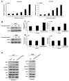
Silencing of Rap1a suppresses melanogenesis. (A,B) Levels of Rap1a mRNA and protein in B16F10 cells treated with milk exosomes were examined by qRT-PCR and Western blotting; n = 3, * p < 0.05, ** p < 0.01. (C) Levels of Rap1a protein were determined by Western blotting in negative control siRNA (NC-siRNA) or Rap1a siRNA-treated cells. (D) B16F10 cells were transfected with NC-siRNA or Rap1a-siRNA. A cell viability assay (WST assay) was performed at 48 h post-transfection. (E,F) Tyrosinase activity and melanin contents were assayed in the cells transfected with NC-siRNA or Rap1a-siRNA; n = 3, ** p < 0.01. (G) Expression of the proteins MITF and TYR was measured in Rap1a siRNA-treated cells by Western blotting.
Figure 7
Figure 7
The Akt-GSK3β signaling pathway is affected by milk exosomes. (A) Protein levels of pAkt, total Akt, pGSK3β and total GSK3β in B16F10 cells transfected with NC-siRNA or Rap1a-siRNA were measured by Western blotting. (B) Akt and GSK3β protein levels were analyzed in cells treated with 20 or 50 μg/mL of milk exosomes. (C) After the cells were transfected with a negative control (NC) or a miR-2478 mimic, Akt and GSK3β protein levels were assayed by Western blotting. (D) In the presence of 50 μg/mL of exosomes, cells were transfected with the NC or miR-2478 inhibitor. (E) Cells exposed to 50 μg/mL of exosomes were treated with the Akt inhibitor GSK 690693. The levels of MITF, TYR and GSK3β in B16F10 were analyzed by Western blot analysis.
Figure 8
Figure 8
Milk exosomes suppress melanogenesis in human melanoma cells and melanocytes. (A) MNT-1 human melanoma cells and NHEM human melanocytes were exposed to 20 or 50 μg/mL of milk exosomes for 48 h, and cell viability was evaluated by a WST assay. (B,C) Tyrosinase activity and melanin contents in the cells treated with milk exosomes were measured; n = 3, ** p < 0.01, *** p < 0.001. (D) Human skin tissues (MelanoDerm) were treated with PBS as a control, or 50 or 100 μg/mL of exosomes, and then photographed. The level of pigmentation in the skin was measured by the L value. (E) Paraffin-embedded tissue sections were stained using hematoxylin and eosin (H&E). The melanin pigment of MelanoDerm was visualized by Fontana–Masson staining (F&M). (F) Melanin contents were determined in the MelanoDerm tissues exposed to milk exosomes; n = 3, * p < 0.05.
Figure 9
Figure 9
miR-2478 inhibits melanogenesis in human melanoma cells and melanocytes. (A) MiR-2478 expression levels in cells treated with milk exosomes were measured by qRT-PCR; n = 3, * p < 0.05, ** p < 0.01, *** p < 0.001. (B) The Rap1a protein level in milk exosome-treated cells was assessed by Western blotting. (C,D) Tyrosinase activity and melanin contents were examined in the negative control (NC) or miR-2478 inhibitor-treated cells with 50 μg/mL of exosomes; n = 3, * p < 0.05. ** p < 0.01. (E) Protein levels of MITF, TYR, pAkt, total Akt, pGSK3β and total GSK3β in NC or miR-2478 inhibitor-transfected cells exposed to milk exosomes were measured by Western blotting.
Figure 10
Figure 10
The proposed model explaining how milk exosomes inhibit melanin production in melanoma cells. In the absence of milk exosomes, Rap1a maintains the active form of GSK3β by inhibiting Akt phosphorylation. The dephosphorylation of GSK3β upregulates the melanogenesis-related protein MITF, which promotes melanin production by driving the expression of tyrosinase. However, in the presence of milk exosomes, miR-2478-carrying milk exosomes inhibit Rap1a expression in melanoma cells, and thus promote the activation of its downstream mediator Akt (via phosphorylation), resulting in GSK3β phosphorylation at Ser9; this change inactivates GSK3β and represses the expression of melanogenesis-related genes such as MITF and TYR, thereby inhibiting melanin production.

References

    1. Draelos Z.D. Cosmeceuticals: What’s real, What’s not. Dermatol. Clin. 2019;37:107–115. doi: 10.1016/j.det.2018.07.001. - DOI - PubMed
    1. Searle T., Al-Niaimi F., Ali F.R. The top 10 cosmeceuticals for facial hyperpigmentation. Dermatol. Ther. 2020;33:e14095. doi: 10.1111/dth.14095. - DOI - PubMed
    1. Slominski R.M., Zmijewski M.A., Slominski A.T. The role of melanin pigment in melanoma. Exp. Dermatol. 2015;24:258–259. doi: 10.1111/exd.12618. - DOI - PMC - PubMed
    1. Praetorius C., Sturm R.A., Steingrimsson E. Sun-induced freckling: Ephelides and solar lentigines. Pigment Cell Melanoma Res. 2014;27:339–350. doi: 10.1111/pcmr.12232. - DOI - PubMed
    1. Serre C., Busuttil V., Botto J.M. Intrinsic and extrinsic regulation of human skin melanogenesis and pigmentation. Int. J. Cosmet. Sci. 2018;40:328–347. doi: 10.1111/ics.12466. - DOI - PubMed

Publication types

MeSH terms

Grants and funding

LinkOut - more resources