Review
. 2021 Oct 26;10(11):2898.
doi: 10.3390/cells10112898.

Molecular Mechanisms of mtDNA-Mediated Inflammation

Affiliations
Review

Molecular Mechanisms of mtDNA-Mediated Inflammation

Anna De Gaetano et al. Cells. .

Abstract

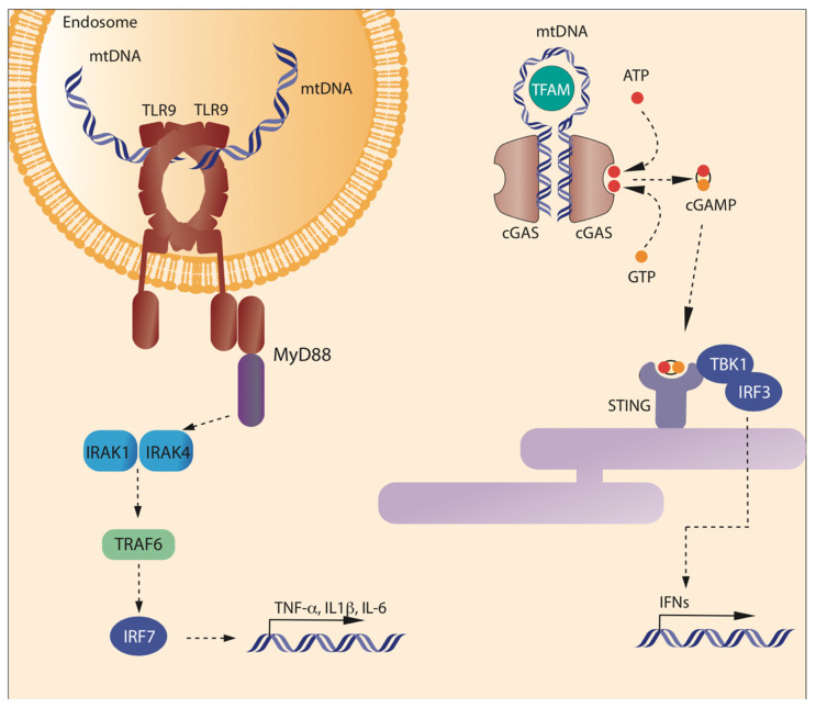
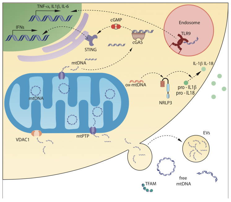
Besides their role in cell metabolism, mitochondria display many other functions. Mitochondrial DNA (mtDNA), the own genome of the organelle, plays an important role in modulating the inflammatory immune response. When released from the mitochondrion to the cytosol, mtDNA is recognized by cGAS, a cGAMP which activates a pathway leading to enhanced expression of type I interferons, and by NLRP3 inflammasome, which promotes the activation of pro-inflammatory cytokines Interleukin-1beta and Interleukin-18. Furthermore, mtDNA can be bound by Toll-like receptor 9 in the endosome and activate a pathway that ultimately leads to the expression of pro-inflammatory cytokines. mtDNA is released in the extracellular space in different forms (free DNA, protein-bound DNA fragments) either as free circulating molecules or encapsulated in extracellular vesicles. In this review, we discussed the latest findings concerning the molecular mechanisms that regulate the release of mtDNA from mitochondria, and the mechanisms that connect mtDNA misplacement to the activation of inflammation in different pathophysiological conditions.

Keywords: STING; TLR9; extracellular cf-mtDNA; inflammasome; mitochondria; mtDNA.


Conflict of interest statement

The authors declare no conflict of interest.

Figures

Figure 1
Figure 1
Binding of mtDNA to TLR9 and cGAS. Left: Toll-like receptor 9 (TLR9) is expressed in the inner face of the endosome membranes, as a homodimeric complex. TLR9 is activated by unmethylated CpG sequences present in DNA molecules, including mtDNA, through a sequence specific binding to the N-term of the C-shaped leucine-rich repeat region of TLR9. Each monomer binds different DNA molecules. Once bound to DNA, the cytosolic domain of TLR9 promotes the activation of MyD88 pathway, which ultimately leads to the transcription of inflammatory cytokines. In the cytosol, mtDNA can be bound by cyclic GMP-AMP Synthase (cGAS) that forms form cyclic GMP-AMP (cGAMP) from GTP and ATP. cGAMP binds to Stimulator of Interferon Genes (STING) on the endoplasmic reticulum. STING promotes the phosphorylation of IRF3 mediated by TBK1, which leads to the transcription of inflammatory genes.
Figure 2
Figure 2
Mechanisms of mtDNA release from mitochondria, and of mtDNA sensing. mtDNA can be released by mitochondria into the cytosol, or in the extracellular space, either as circular molecules, DNA fragments, or DNA associated with mitochondrial proteins. Leakage from mDNA can be mediated by mitochondrial Permeability Transition Pore (mPTP), that causes swelling of the mitochondrial inner chamber and loss of impermeability of the Mitochondrial Inner Membrane (MIM), and by pores on the Mitochondrial Outer Membrane (MOM), formed by pro-apoptotic protein BAX or by the oligomers of the mitochondrial porin voltage-dependent anion-selective channel 1 (VDAC1). Once in the cytosol, mtDNA can interact with cGAS, which activates a pathway leading to enhanced transcription of type 1 interferons, or with NLRP3, particularly when oxidized. NLRP3 inflammasomes activate IL-1β and IL-18 by post-translational cleavage. Cells can also take up mtDNA from surrounding environment by phagocytosis; once in the endosomes, mtDNA can interact with the Toll-like receptor 9, which activates a pathway leading to transcription of pro-inflammatory cytokines TNF-α, IL-1β, and IL-6. Finally, mtDNA can be released outside the cells either by passive release, mediated by rupture of plasma cell membrane integrity, or by active release through extracellular vesicles.

References

    1. Buck M.D., Sowell R.T., Kaech S.M., Pearce E.L. Metabolic Instruction of Immunity. Cell. 2017;169:570–586. doi: 10.1016/j.cell.2017.04.004. - DOI - PMC - PubMed
    1. Rambold A.S., Pearce E.L. Mitochondrial Dynamics at the Interface of Immune Cell Metabolism and Function. Trends Immunol. 2018;39:6–18. doi: 10.1016/j.it.2017.08.006. - DOI - PubMed
    1. Dela Cruz C.S., Kang M.J. Mitochondrial dysfunction and damage associated molecular patterns (DAMPs) in chronic inflammatory diseases. Mitochondrion. 2018;41:37–44. doi: 10.1016/j.mito.2017.12.001. - DOI - PMC - PubMed
    1. Roger A.J., Munoz-Gomez S.A., Kamikawa R. The Origin and Diversification of Mitochondria. Curr. Biol. 2017;27:R1177–R1192. doi: 10.1016/j.cub.2017.09.015. - DOI - PubMed
    1. Sagan L. On the origin of mitosing cells. J. Theor. Biol. 1967;14:255–274. doi: 10.1016/0022-5193(67)90079-3. - DOI - PubMed

Publication types