Review
. 2021 Oct 29;10(11):2945.
doi: 10.3390/cells10112945.

Mitochondria in Diabetic Kidney Disease

Affiliations
Review

Mitochondria in Diabetic Kidney Disease

Amna Ayesha Ahmad et al. Cells. .

Abstract

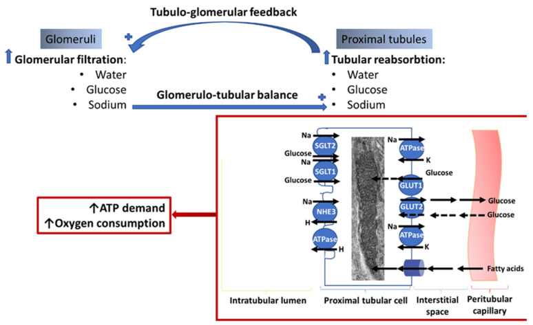
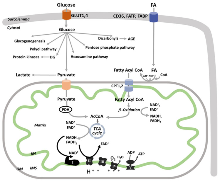
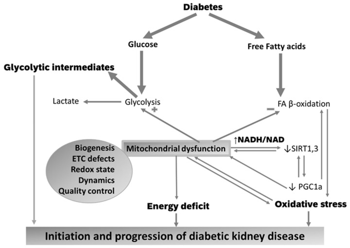
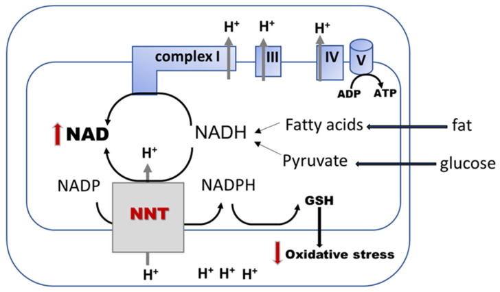
Diabetic kidney disease (DKD) is the leading cause of end stage renal disease (ESRD) in the USA. The pathogenesis of DKD is multifactorial and involves activation of multiple signaling pathways with merging outcomes including thickening of the basement membrane, podocyte loss, mesangial expansion, tubular atrophy, and interstitial inflammation and fibrosis. The glomerulo-tubular balance and tubule-glomerular feedback support an increased glomerular filtration and tubular reabsorption, with the latter relying heavily on ATP and increasing the energy demand. There is evidence that alterations in mitochondrial bioenergetics in kidney cells lead to these pathologic changes and contribute to the progression of DKD towards ESRD. This review will focus on the dialogue between alterations in bioenergetics in glomerular and tubular cells and its role in the development of DKD. Alterations in energy substrate selection, electron transport chain, ATP generation, oxidative stress, redox status, protein posttranslational modifications, mitochondrial dynamics, and quality control will be discussed. Understanding the role of bioenergetics in the progression of diabetic DKD may provide novel therapeutic approaches to delay its progression to ESRD.

Keywords: bioenergetics; diabetes; diabetic kidney disease; mitochondria; redox status.


Conflict of interest statement

The authors declare no conflict of interest.

Figures

Figure 1
Figure 1
The crosstalk between glomeruli and proximal tubules is an energy-consuming process. The increased proximal tubular reabsorption of components within the glomerular filtrate involves apical transporters including Sodium Glucose Transporters (SGLT) 1 and 2, Sodium Hydrogen Exchanger 3 (NHE3), and Hydrogen-ATPase. The gradients needed for the apical transport are provided by the Na-K ATPase. Glucose exits via basal GLUT2 to enter the peritubular capillaries. Hyperglycemia either reverse or impede the glucose transport via GLUT2 and facilitate the glucose uptake via GLUT1, thus favoring intracellular glucose accumulation. The major ATP source is fatty acid (FA) β-oxidation within the mitochondria; however, glucose can also be used for energy generation although the bulk is only in transit to be recovered from the urinary space to the blood.
Figure 2
Figure 2
Metabolism of energetic substrates in kidney cells. The basal and insulin-dependent glucose uptake into kidney cells is mediated by the Glucose Transporters (GLUT) 1 and 4. Free fatty acids (FA) enters kidney cells by a route supported by the protein CD36, FA transport proteins (FATPs) [69], and FA binding proteins (FABPs) [70]. FA uptake by kidney cells is not hormonally regulated and driven by their circulating availability. Glucose follows multiple metabolic pathways including glycolysis, glycogen synthesis (glycogenogenesis), polyol pathway (with sorbitol and fructose formation), conversion to either diacylglycerol (DAG) to activate protein kinases or to dicarbonyls (i.e., methylglyoxal) to form advanced glycation end products (AGE), and is shuttled into the hexosamine biosynthetic or pentose phosphate pathways. Pyruvate is either converted to lactate (aerobic glycolysis [71]) or transported into mitochondria via a mitochondrial pyruvate carrier to be converted by pyruvate dehydrogenase (PDH) to acetyl-CoA (AcCoA) for the tricarboxylic acid (TCA) cycle. For simplicity, extramitochondrial glucose fluxes are not shown in detail. After entry into the cell, long chain FAs are activated to FA-CoA that is either esterified as triacylglycerol (stored in the cytosol, not shown) or enter the mitochondria via carnitine palmitoyltransferases (CPT1 and 2) to be oxidized. The end products of each FA β-oxidation cycle are NADH, FADH2, and acetyl-CoA, which are further oxidized by the electron transport chain (ETC) complexes or TCA, respectively, ultimately leading to ATP synthesis via mitochondrial oxidative phosphorylation. Mitochondrial oxidative phosphorylation is the main ATP provider. While electrons are transferred from the reducing equivalents, NADH and FADH2, to oxygen by the ETC complexes, an electrochemical gradient is built across the mitochondrial inner membrane (IM), which is used by the ATP synthase (complex V) to form ATP. OM: outer membrane; IMS: intermembrane space.
Figure 3
Figure 3
Excessive energetic substrates in diabetes leads to mitochondrial defects that cause reductive stress, oxidative stress, and energy deficit. Decreased insulin activity in diabetes provides an excess of circulating energetic substrates (i.e., glucose, free fatty acids) that enter the kidney cells by an unregulated route and are used in metabolism. An increase in the glycolytic flux that is not matched by an active mitochondrial oxidative phosphorylation leads to an accumulation of glycolytic intermediates that follow alternative metabolic pathway in the cytosol, which are responsible for inflammation, fibrosis, and epigenetic changes. The increased fatty acid (FA) oxidation observed in early diabetes, which is driven by PGC1α overexpression, leads to increased mitochondrial oxidative stress. During more advanced diabetes, mitochondrial defects limit FA oxidation and increase the NADH/NAD redox ratio (reductive stress) that restrains the mitochondrial lysine deacetylase, sirtuin 3 (SIRT3) activity. SIRT3 deacetylates and activates FA oxidation enzymes, ETC components, and PGC1α. Mitochondrial dysfunction has multiple pathogenic mechanisms including decreased PGC1α -mediated mitochondrial biogenesis and posttranslational modifications of mitochondrial proteins (oxidation, lysine acetylation).
Figure 4
Figure 4
The role of nicotinamide nucleotide transhydrogenase (NNT) in decreasing the reductive and oxidative stress. NNT is an inner mitochondrial membrane enzyme that transfers electrons from mitochondrial NADH to NADPH by using the inner membrane proton motive force to boost the peroxide antioxidant defense, such as reduced glutathione (GSH) [186,187]. The enzyme reduces NADPH by oxidizing NADH that is generated by oxidizing energetic substrates (i.e., fat and glucose), thus decreasing the reductive stress (NADH/NAD ratio).

References

    1. Han Q., Zhu H., Chen X., Liu Z. Non-genetic mechanisms of diabetic nephropathy. Front. Med. 2017;11:319–332. doi: 10.1007/s11684-017-0569-9. - DOI - PubMed
    1. Alicic R.Z., Rooney M.T., Tuttle K.R. Diabetic Kidney Disease: Challenges, Progress, and Possibilities. Clin. J. Am. Soc. Nephrol. 2017;12:2032–2045. doi: 10.2215/CJN.11491116. - DOI - PMC - PubMed
    1. Pugliese G., Penno G., Natali A., Barutta F., Di Paolo S., Reboldi G., Gesualdo L., De Nicola L., Italian Diabetes Society. the Italian Society of Nephrology Diabetic kidney disease: New clinical and therapeutic issues. Joint position statement of the Italian Diabetes Society and the Italian Society of Nephrology on “The natural history of diabetic kidney disease and treatment of hyperglycemia in patients with type 2 diabetes and impaired renal function”. Nutr. Metab. Cardiovasc. Dis. 2019;29:1127–1150. doi: 10.1016/j.numecd.2019.07.017. - DOI - PubMed
    1. Afkarian M., Zelnick L.R., Hall Y.N., Heagerty P.J., Tuttle K., Weiss N.S., de Boer I.H. Clinical Manifestations of Kidney Disease Among US Adults with Diabetes, 1988–2014. JAMA. 2016;316:602–610. doi: 10.1001/jama.2016.10924. - DOI - PMC - PubMed
    1. Minutolo R., Gabbai F.B., Provenzano M., Chiodini P., Borrelli S., Garofalo C., Sasso F.C., Santoro D., Bellizzi V., Conte G., et al. Cardiorenal prognosis by residual proteinuria level in diabetic chronic kidney disease: Pooled analysis of four cohort studies. Nephrol. Dial. Transplant. 2018;33:1942–1949. doi: 10.1093/ndt/gfy032. - DOI - PubMed

Publication types