Review
. 2021 Nov 6;10(11):3053.
doi: 10.3390/cells10113053.

TRPV4 Mechanotransduction in Fibrosis

Affiliations
Review

TRPV4 Mechanotransduction in Fibrosis

Ravi K Adapala et al. Cells. .

Abstract

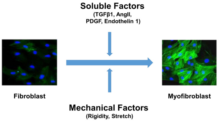
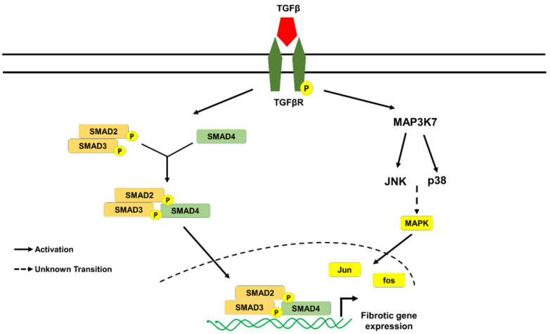
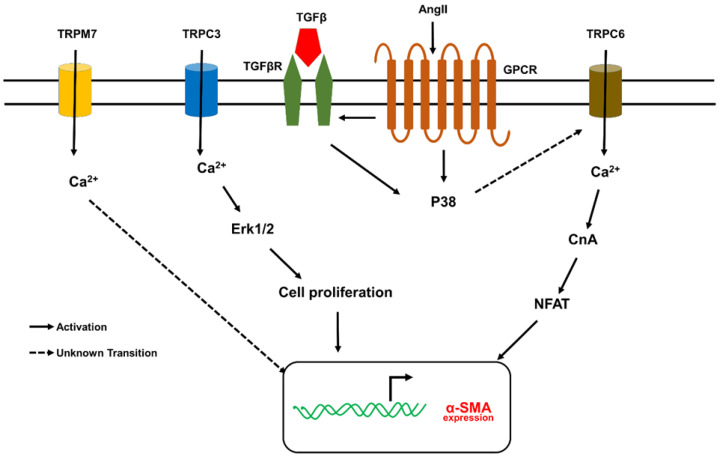
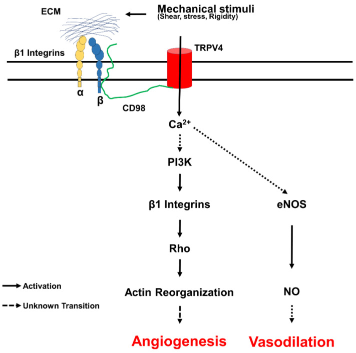
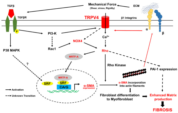
Fibrosis is an irreversible, debilitating condition marked by the excessive production of extracellular matrix and tissue scarring that eventually results in organ failure and disease. Differentiation of fibroblasts to hypersecretory myofibroblasts is the key event in fibrosis. Although both soluble and mechanical factors are implicated in fibroblast differentiation, much of the focus is on TGF-β signaling, but to date, there are no specific drugs available for the treatment of fibrosis. In this review, we describe the role for TRPV4 mechanotransduction in cardiac and lung fibrosis, and we propose TRPV4 as an alternative therapeutic target for fibrosis.

Keywords: TGF-β; TRPV4; calcium; extracellular matrix; fibroblast; fibrosis; mechanotransduction; myofibroblast.


Conflict of interest statement

The authors declare no conflict of interest.

Figures

Figure 1
Figure 1
Fibroblast differentiation into myofibroblasts is a key event in fibrosis. Both soluble factors (TGF-β or Ang II or PDGF or Endothelin 1) and mechanical factors (stretch and stiffness) are required for cardiac fibroblasts differentiation into hyper-secretory and hyper-contractile phenotype myofibroblasts (α-SMA (green) expression and incorporation into stress fibers are known markers of myofibroblasts; nuclei were stained in blue). Ang II = angiotensin II; α-SMA = α-smooth muscle actin; TGF-β = transforming growth factor β. PDGF = Platelet derived growth factor.
Figure 2
Figure 2
TGF-β signaling fibroblasts differentiation into myofibroblasts. TGF-β is the major soluble factor implicated in fibroblast differentiation. The canonical pathway includes binding of TGF-β to the TGF-β receptors resulting in the activation of SMAD2/3, which forms a complex with SMAD4, translocates to nucleus, and induces fibrotic gene expression. In contrast, non-canonical pathway includes p38 MAPK/JNK pathway, which activates Jun/Fos-dependent fibrotic gene expression. α-SMA = α-smooth muscle actin; TGF-β = transforming growth factor β; TGFβR = transforming growth factor β receptor.
Figure 3
Figure 3
Schematic showing possible mechanisms by which TRP channels (other than TRPV4) regulate fibroblast differentiation. TRPC3, TRPC6, and TRPM7 have been implicated in fibroblast (atrial and ventricular) differentiation. TGF-β shown to influence the expression/activity of these three TRP channels. TRPC3 signaling demonstrated to be critical for atrial fibroblast proliferation, migration, and fibrosis. The possible signaling mechanism appears to be AngII/AT1R/TGF-β1 mediated increase in TRPC3 expression via NFAT dependent downregulation of miR-26, which in turn activates fibroblast proliferation through ERK1/2. TGF-β was also shown to increase the expression of TRPC6 channels via p38/SRF and then calcium influx from TRPC6 induces fibroblast differentiation through CnA/NFAT signaling. TRPM7 shown to regulate mechanical stretch-induced fibroblast differentiation (atrial and adventitial) though p38 MAPK/JNK pathway. Ang II, angiotensin II; CnA, calcineurin; miR-26, microRNA26; NFAT, Nuclear factor of activated T-cells; ROCK, Rho associated protein kinase; SRF, serum responsive factor; α-SMA, α-smooth muscle actin; TGF-β, transforming growth factor β.
Figure 4
Figure 4
TRPV4 mechanotransduction in endothelial cells. Cells sense mechanical forces exerted on extracellular matrix through integrins in focal adhesion. Application of mechanical force through integrins results in ultra-rapid activation of TRPV4 channels via CD98 proteins. TRPV4 mediated calcium influx activates additional integrins through PI3 Kinase, which induces transient downregulation and later stabilization of Rho facilitating cytoskeletal reorientation and directed migration of EC required for physiological angiogenesis. TRPV4-mediated calcium influx can activate endothelial nitric oxide synthase (eNOS) and generation of nitric oxide (NO), which induces vasodilation.
Figure 5
Figure 5
TRPV4 integrates soluble and mechanical signaling during fibroblast differentiation to myofibroblast. TRPV4 senses mechanical forces and activates Rho/Rho kinase pathway. Rho/Rho kinase dependent polymerization of actin releases MRTF-A from actin monomers, which then translocates into nucleus and induces α-SMA expression by binding with CArG sequences together with SRF. Rho/Rho kinase, on the other hand, enables α-SMA incorporation into the stress fibers, resulting in fibroblast differentiation into myofibroblasts. TGF-β activates PI3K-gamma, which forms complex with TRPV4; the complex is translocated to the plasma membrane and activates calcium influx at the Rho/MRTF-A pathway. TRPV4/TGF-β was also shown to activate NOX4 via PI3K/Rac1, which feeds into Rho pathway. In addition to inducing fibroblast differentiation to myofibroblasts and fibrotic gene expression, Rho/Rho kinase pathway activates PAI-1 expression and inhibits matrix degradation. Myofibroblasts are hypercontractile and apply tensional forces on the ECM and activate integrins, resulting in the release of TGF-β from latent complex inducing a positive feedback loop and enhancing fibroblast differentiation to myofibroblasts, excessive ECM production, and eventually fibrosis. Since, TRPV4 integrates soluble (TGF-β) and mechanical (integrin/Rho) signaling required for fibroblast differentiation and fibrosis, we propose TRPV4 as the novel therapeutic target for fibrosis.

References

    1. Horn M., Trafford A.W. Aging and the cardiac collagen matrix: Novel mediators of fibrotic remodelling. J. Mol. Cell. Cardiol. 2016;93:175–185. doi: 10.1016/j.yjmcc.2015.11.005. - DOI - PMC - PubMed
    1. Talman V., Ruskoaho H. Cardiac fibrosis in myocardial infarction—from repair and remodeling to regeneration. Cell Tissue Res. 2016;365:563–581. doi: 10.1007/s00441-016-2431-9. - DOI - PMC - PubMed
    1. DeLeon-Pennell K.Y., Meschiari C.A., Jung M., Lindsey M.L. Matrix Metalloproteinases in Myocardial Infarction and Heart Failure. Prog. Mol. Biol. Transl. Sci. 2017;147:75–100. doi: 10.1016/bs.pmbts.2017.02.001. - DOI - PMC - PubMed
    1. Frangogiannis N.G. The Extracellular Matrix in Ischemic and Nonischemic Heart Failure. Circ. Res. 2019;125:117–146. doi: 10.1161/CIRCRESAHA.119.311148. - DOI - PMC - PubMed
    1. Fan Z., Guan J. Antifibrotic therapies to control cardiac fibrosis. Biomater. Res. 2016;20:13. doi: 10.1186/s40824-016-0060-8. - DOI - PMC - PubMed

Publication types

Substances

LinkOut - more resources