

The New Kid on the Block: HLA-C, a Key Regulator of Natural Killer Cells in Viral Immunity
- PMID: 34831331
- PMCID: PMC8620871
- DOI: 10.3390/cells10113108
The New Kid on the Block: HLA-C, a Key Regulator of Natural Killer Cells in Viral Immunity
Abstract
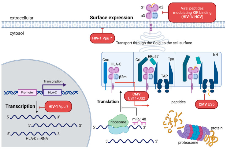
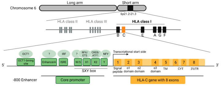
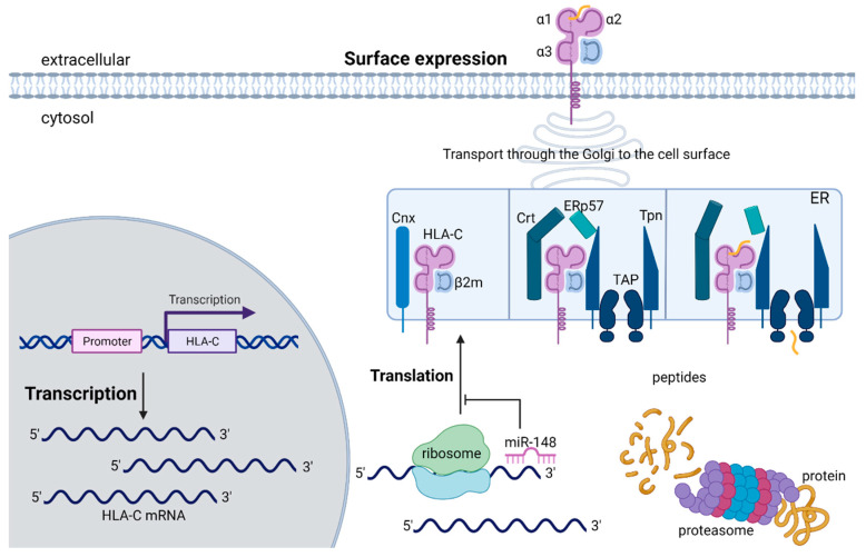
The human leukocyte antigen system (HLA) is a cluster of highly polymorphic genes essential for the proper function of the immune system, and it has been associated with a wide range of diseases. HLA class I molecules present intracellular host- and pathogen-derived peptides to effector cells of the immune system, inducing immune tolerance in healthy conditions or triggering effective immune responses in pathological situations. HLA-C is the most recently evolved HLA class I molecule, only present in humans and great apes. Differentiating from its older siblings, HLA-A and HLA-B, HLA-C exhibits distinctive features in its expression and interaction partners. HLA-C serves as a natural ligand for multiple members of the killer-cell immunoglobulin-like receptor (KIR) family, which are predominately expressed by natural killer (NK) cells. NK cells are crucial for the early control of viral infections and accumulating evidence indicates that interactions between HLA-C and its respective KIR receptors determine the outcome and progression of viral infections. In this review, we focus on the unique role of HLA-C in regulating NK cell functions and its consequences in the setting of viral infections.
Keywords: HLA-C; NK cells; killercell immunoglobulin-like receptors; viral infection.
Conflict of interest statement
The authors declare no conflict of interests.
Figures

Similar articles
-
HLA Class I Downregulation by HIV-1 Variants from Subtype C Transmission Pairs.J Virol. 2018 Mar 14;92(7):e01633-17. doi: 10.1128/JVI.01633-17. Print 2018 Apr 1. J Virol. 2018. PMID: 29321314 Free PMC article.
-
Hierarchy of the human natural killer cell response is determined by class and quantity of inhibitory receptors for self-HLA-B and HLA-C ligands.J Immunol. 2007 Nov 1;179(9):5977-89. doi: 10.4049/jimmunol.179.9.5977. J Immunol. 2007. PMID: 17947671
-
Killer cell immunoglobulin-like receptor genotype and killer cell immunoglobulin-like receptor-human leukocyte antigen C ligand compatibility affect the severity of hepatitis C virus recurrence after liver transplantation.Liver Transpl. 2009 Apr;15(4):390-9. doi: 10.1002/lt.21673. Liver Transpl. 2009. PMID: 19326408
-
Why natural killer cells are not enough: a further understanding of killer immunoglobulin-like receptor and human leukocyte antigen.Fertil Steril. 2017 Jun;107(6):1273-1278. doi: 10.1016/j.fertnstert.2017.04.018. Epub 2017 May 10. Fertil Steril. 2017. PMID: 28501365 Review.
-
Natural killer cell recognition of HLA class I molecules.Rev Immunogenet. 2000;2(3):433-48. Rev Immunogenet. 2000. PMID: 11256749 Review.
Cited by
-
Natural Killer (NK) Cell Alloreactivity in Haploidentical Stem Cell Transplantation.Cells. 2025 Jul 16;14(14):1091. doi: 10.3390/cells14141091. Cells. 2025. PMID: 40710344 Free PMC article. Review.
-
Coordinated Loss and Acquisition of NK Cell Surface Markers Accompanied by Generalized Cytokine Dysregulation in COVID-19.Int J Mol Sci. 2023 Jan 19;24(3):1996. doi: 10.3390/ijms24031996. Int J Mol Sci. 2023. PMID: 36768315 Free PMC article.
-
Hypoimmunogenic CD19 CAR-NK cells derived from embryonic stem cells suppress the progression of human B-cell malignancies in xenograft animals.Front Immunol. 2024 Nov 27;15:1504459. doi: 10.3389/fimmu.2024.1504459. eCollection 2024. Front Immunol. 2024. PMID: 39664387 Free PMC article.
-
Immunogenetic profiles of 9 human herpes virus envelope glycoproteins.Sci Rep. 2024 Sep 9;14(1):20924. doi: 10.1038/s41598-024-71558-1. Sci Rep. 2024. PMID: 39251790 Free PMC article.
-
Immunogenomics of Killer Cell Immunoglobulin-Like Receptor (KIR) and HLA Class I: Coevolution and Consequences for Human Health.J Allergy Clin Immunol Pract. 2022 Jul;10(7):1763-1775. doi: 10.1016/j.jaip.2022.04.036. Epub 2022 May 10. J Allergy Clin Immunol Pract. 2022. PMID: 35561968 Free PMC article.
References
-
- Peakman M., Vergani D. Basic and Clinical Immunology. Churchill Livingstone; London, UK: 2009.
Publication types
MeSH terms
Substances
Grants and funding
LinkOut - more resources
Full Text Sources
Research Materials
