Review
. 2021 Nov 13;10(11):3162.
doi: 10.3390/cells10113162.

Mechanisms of Plasticity in Subcortical Visual Areas

Affiliations
Review

Mechanisms of Plasticity in Subcortical Visual Areas

Maël Duménieu et al. Cells. .

Abstract

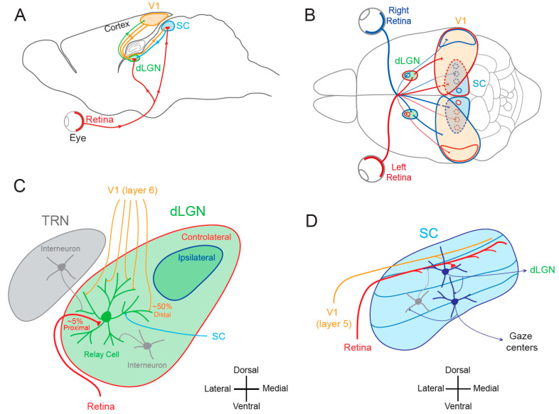
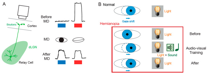
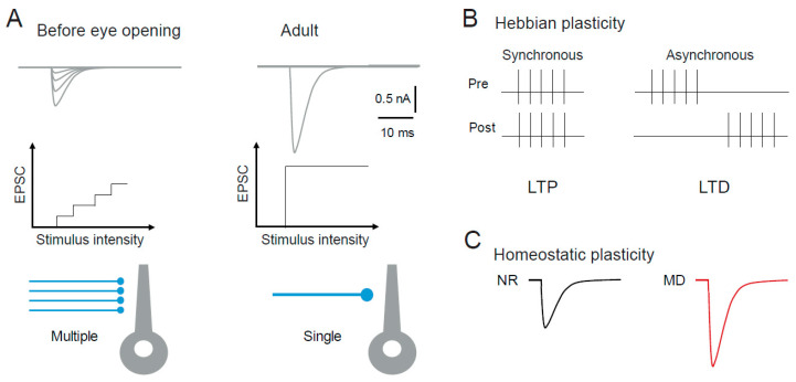
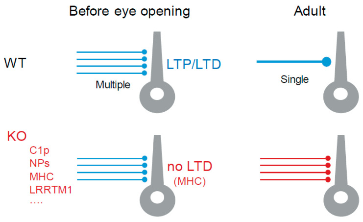
Visual plasticity is classically considered to occur essentially in the primary and secondary cortical areas. Subcortical visual areas such as the dorsal lateral geniculate nucleus (dLGN) or the superior colliculus (SC) have long been held as basic structures responsible for a stable and defined function. In this model, the dLGN was considered as a relay of visual information travelling from the retina to cortical areas and the SC as a sensory integrator orienting body movements towards visual targets. However, recent findings suggest that both dLGN and SC neurons express functional plasticity, adding unexplored layers of complexity to their previously attributed functions. The existence of neuronal plasticity at the level of visual subcortical areas redefines our approach of the visual system. The aim of this paper is therefore to review the cellular and molecular mechanisms for activity-dependent plasticity of both synaptic transmission and cellular properties in subcortical visual areas.

Keywords: Hebbian plasticity; homeostatic plasticity; intrinsic plasticity; lateral geniculate nucleus; superior colliculus; synaptic plasticity; visual system.


Conflict of interest statement

The authors declare no conflict of interest.

Figures

Figure 1
Figure 1
Visual pathways. (A) Sagittal view of the rodent visual system. V1, primary visual area; SC, superior colliculus; dLGN, dorsal lateral geniculate nucleus. (B) Superior view of the rodent visual system. Red, visual inputs from the left eye. Blue, visual inputs from the right eye. (C) Simplified synaptic organization of visual inputs to rodent dLGN. The relay cell receives 3 types of excitatory inputs: (1) small amount (~5%) of functionally powerful contralateral inputs from the retina on proximal dendrite (red), (2) numerous (~50%) but functionally weak feed-back inputs from V1 (orange) on distal dendrites and (3) from the SC (light blue) on medial and distal dendrites. In addition, it is inhibited by interneurons located in the TRN (thalamic reticular nucleus) and in the dLGN (grey). (D) Principal inputs and outputs of rodent SC neurons. In the superficial layer, SC neurons receive excitatory inputs from the retina (red) and from V1 (orange) and an inhibitory feed-back (grey) from interneurons located deeper in the SC. Superficial excitatory neurons contact deeper premotor neurons and neurons in the dLGN. Premotor neurons in the deep layer feed gaze centers of the brain. Adapted from [11].
Figure 2
Figure 2
Functional plasticity in subcortical visual areas. (A) In the dLGN. Left, calcium imaging setup in presynaptic boutons of dLGN relay cell. Right, calcium signals evoked by visual stimulation (blue bar, stimulation of the right eye; red bar, stimulation of the left eye). Upper part, before MD; middle part, MD; lower part, after MD. Visual response is evoked only through one eye before MD, whereas visual response is evoked through each eye after MD on the left eye (adapted from [38]). (B) In the SC. In normal patients, gaze orientation occurs upon visual stimulation (light). In hemianopia patients, the same light stimulus produces no gaze shift. During audio-visual training where light is associated with a sound localized in the same region of space, gaze shift occurs. After training, light alone produced a gaze shift (adapted from [41,42]).
Figure 3
Figure 3
Plasticity in the dLGN. (A) Structural plasticity. Refinement, synapse elimination and synapse strengthening at rodent retino-geniculate inputs during postnatal development. Synaptic currents evoked by increasing stimulus intensity before eye opening (left) and in the adult (right). Note the multiple and small synaptic responses in immature dLGN neurons and the all-or-none and large response in mature dLGN neurons. (B) Hebbian synaptic plasticity at retino-geniculate synapses induced by pairing presynaptic stimulation with postsynaptic firing with synchronous (left) or asynchronous (right) relation. Adapted from [62]. (C) Homeostatic plasticity at cortico-geniculate synapses. MD induces an up-regulation of synaptic transmission. Adapted from [63]. NR, normal rearing.
Figure 4
Figure 4
Molecular mechanisms of plasticity in subcortical visual areas. Top, in WT animals before eye opening, thalamocortical neurons are fed by multiple and weak retinal inputs, while in WT adult animals retinal inputs are strong and reduced (refinement). Note that LTP and LTD can be induced at an immature stage. Bottom, in KO animals for proteins involved in synaptic pruning (C1p, NPS, MHC, LRRTM1, and others), no synaptic refinement occurs. Note that LTD is prevented in MHC KO.

References

    1. Weyand T.G. The Multifunctional Lateral Geniculate Nucleus. Rev. Neurosci. 2016;27:135–157. doi: 10.1515/revneuro-2015-0018. - DOI - PubMed
    1. Sherman S.M. Thalamus Plays a Central Role in Ongoing Cortical Functioning. Nat. Neurosci. 2016;19:533–541. doi: 10.1038/nn.4269. - DOI - PubMed
    1. Ghodrati M., Khaligh-Razavi S.-M., Lehky S.R. Towards Building a More Complex View of the Lateral Geniculate Nucleus: Recent Advances in Understanding Its Role. Prog. Neurobiol. 2017;156:214–255. doi: 10.1016/j.pneurobio.2017.06.002. - DOI - PubMed
    1. Basso M.A., Bickford M.E., Cang J. Unraveling Circuits of Visual Perception and Cognition through the Superior Colliculus. Neuron. 2021;109:918–937. doi: 10.1016/j.neuron.2021.01.013. - DOI - PMC - PubMed
    1. Isa T., Marquez-Legorreta E., Grillner S., Scott E.K. The Tectum/Superior Colliculus as the Vertebrate Solution for Spatial Sensory Integration and Action. Curr. Biol. 2021;31:R741–R762. doi: 10.1016/j.cub.2021.04.001. - DOI - PMC - PubMed

Publication types

LinkOut - more resources