. 2021 Nov 17;10(11):3210.
doi: 10.3390/cells10113210.

Therapeutic Benefit of Galectin-1: Beyond Membrane Repair, a Multifaceted Approach to LGMD2B

Affiliations

Therapeutic Benefit of Galectin-1: Beyond Membrane Repair, a Multifaceted Approach to LGMD2B

Mary L Vallecillo-Zúniga et al. Cells. .

Abstract

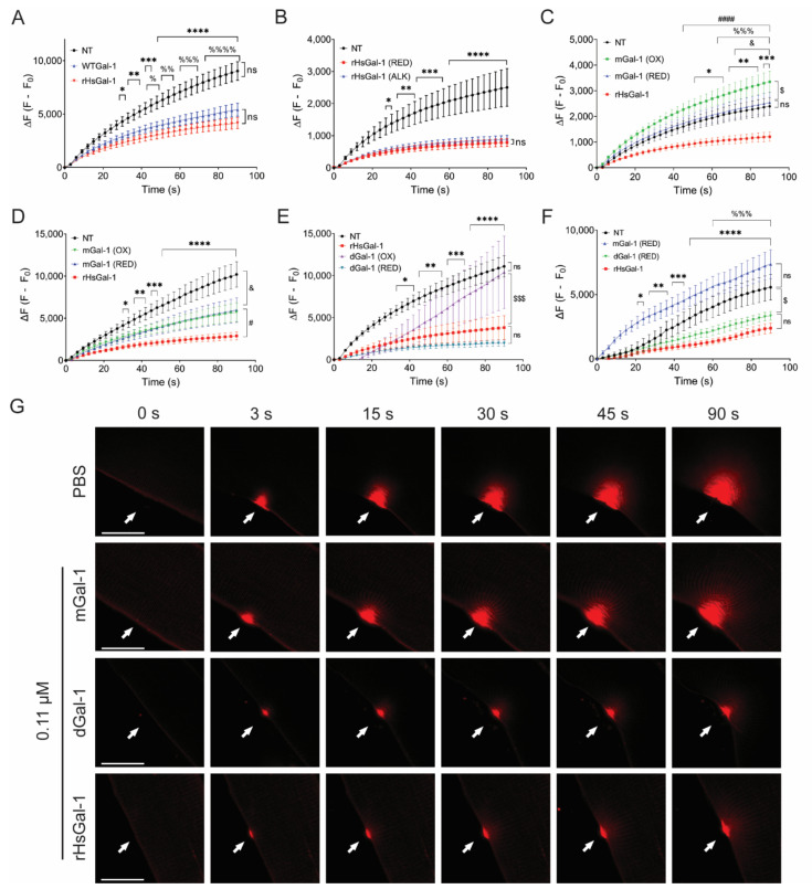
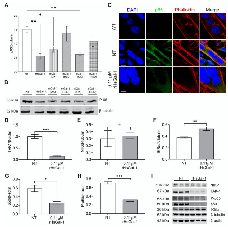
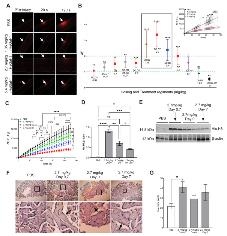
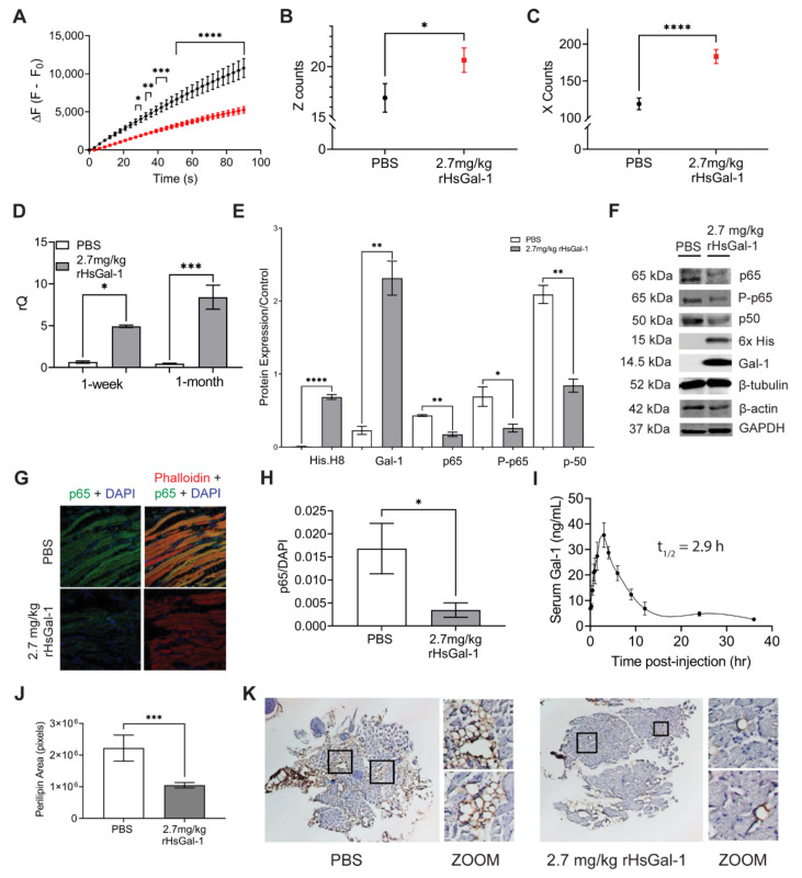
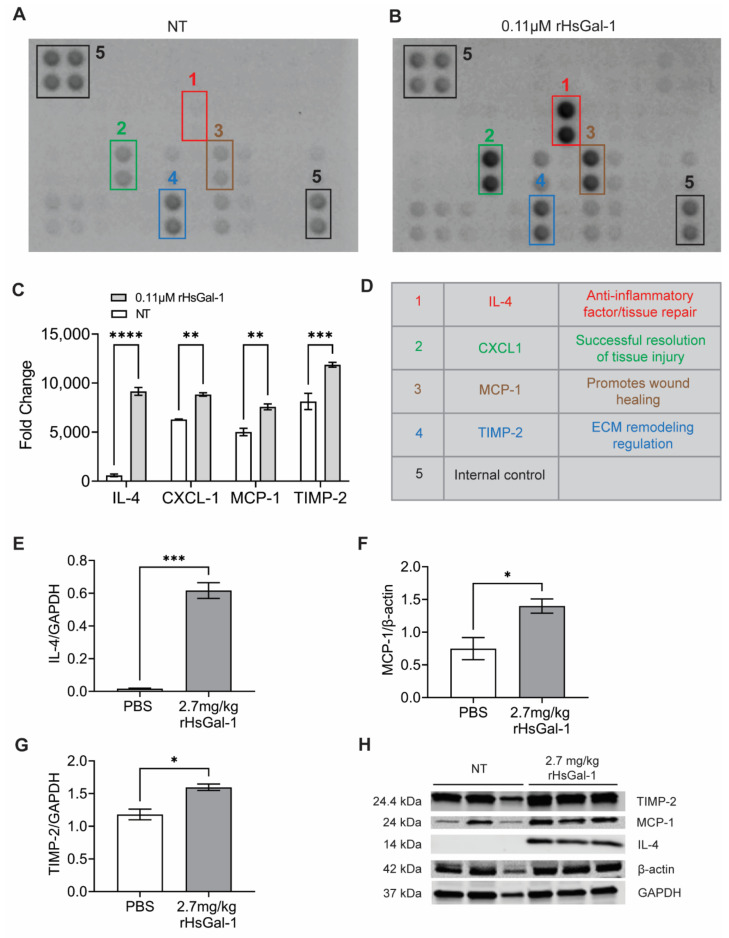
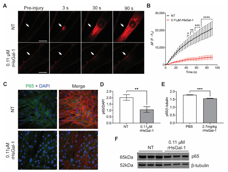
Two of the main pathologies characterizing dysferlinopathies are disrupted muscle membrane repair and chronic inflammation, which lead to symptoms of muscle weakness and wasting. Here, we used recombinant human Galectin-1 (rHsGal-1) as a therapeutic for LGMD2B mouse and human models. Various redox and multimerization states of Gal-1 show that rHsGal-1 is the most effective form in both increasing muscle repair and decreasing inflammation, due to its monomer-dimer equilibrium. Dose-response testing shows an effective 25-fold safety profile between 0.54 and 13.5 mg/kg rHsGal-1 in Bla/J mice. Mice treated weekly with rHsGal-1 showed downregulation of canonical NF-κB inflammation markers, decreased muscle fat deposition, upregulated anti-inflammatory cytokines, increased membrane repair, and increased functional movement compared to non-treated mice. Gal-1 treatment also resulted in a positive self-upregulation loop of increased endogenous Gal-1 expression independent of NF-κB activation. A similar reduction in disease pathologies in patient-derived human cells demonstrates the therapeutic potential of Gal-1 in LGMD2B patients.

Keywords: Galectin-1; LGMD2B; NF-ĸB; cytokines; inflammation; membrane repair; muscular dystrophy.


Conflict of interest statement

Authors of this manuscript have the following declaration of interests: The University of Nevada-Reno has been issued a patent in the U.S. (# US20130065242 A1) and Australia (# 45557BOA/VPB) for “Methods for diagnosing, prognosing and treating muscular dystrophy”. PMVR is an inventor on these patents. Strykagen currently holds the license for this technology. Brigham Young University has a patent for “Galectin-1 immunomodulation and myogenic improvements in muscle diseases and autoimmune disorders.” (#U.S. Pat. No. 62/161,027. PCT/US2021/026232). This does not alter our adherence to MDPI Cells’ policies on sharing data and materials.

Figures

Figure 1
Figure 1
rHsGal-1 is the most efficient type of Gal-1 in helping improve sarcolemmal repair in A/J myotubes and Bla/J myofibers. (A) Quantification of the change in fluorescent intensity inside A/J−/−myotubes following laser injury when treated with 0.11 μM WT Gal-1 and 0.11 μM rHsGal-1 for 48 h compared to NT A/J−/− myotubes. *, = NT vs. 0.11 μM rHsGal-1. % = NT vs. 0.11 μM WT Gal-1. (B) Change in the fluorescent intensity in A/J−/− myotubes following laser injury when treated with 0.11 μM rHsGal-1 and 0.11 μM alkylated rHsGal-1 compared to NT A/J−/− myotubes. (C) Quantified laser injury assay displaying membrane repair differences between 10 min treatment of A/J−/− myotubes with 0.11 μM mGal-1 (oxidized), 0.11 μM mGal-1 (reduced), and 0.11 μM rHsGal-1 compared to NT. $, = mGal-1 (oxidized) vs. mGal-1 (reduced), % = mGal-1 (reduced) vs. rHsGal-1, # = mGal-1 (oxidized) vs. rHsGal-1, & = NT vs. mGal-1 (oxidized), * = NT vs. rHsGal-1. (D) Quantified laser injury assay displaying membrane repair differences between 48 h treatment of A/J−/− myotubes with 0.11 μM mGal-1 (oxidized), 0.11 μM mGal-1 (reduced), and 0.11 μM rHsGal-1 compared to NT. # = mGal-1 (oxidized and reduced) vs. rHsGal-1, & = NT vs. mGal-1 (oxidized and reduced), * = NT vs. rHsGal-1. (E) Quantified laser injury assay displaying membrane repair differences between 48 h treatment of A/J−/− myotubes with 0.11 μM dGal-1 (oxidized), 0.11 μM dGal-1 (reduced), and 0.11 μM rHsGal-1 compared to NT. $ = dGal-1 (oxidized) vs. rHsGal-1, * = NT vs. rHsGal-1 and NT vs. dGal-1 (reduced) and dGal-1 (oxidized) vs. dGal-1 (reduced). (F) Quantified laser injury assay displaying membrane repair differences between 2 h treatment of isolated Bla/J mouse myofibers with 0.11 μM mGal-1 (reduced), 0.11 μM dGal-1 (reduced), and 0.11 μM rHsGal-1 compared to PBS. $ = PBS vs. dGal-1 (reduced), % = PBS vs. rHsGal-1, * = rHsGal-1 and dGal-1 (reduced) vs. mGal-1 (reduced). (G) Representative images of treated explant Bla/J myofibers during laser injury assay taken at 0 s, 3 s, 15 s, 30 s, 45 s, and 90 s with arrows indicating location of injury. All p-values were calculated by Tukey’s multiple comparison test and indicated by ****, %%%%, and #### = p < 0.0001; ***, %%%, and ### = p < 0.001; **, %%, and ## = p < 0.01; and *, %, &, and # = p < 0.05. Scale bars = 20 μm. Error bars represent SEM. n ≥ 23 from 2 independent experiments for each group.
Figure 2
Figure 2
In vitro treatment with rHsGal-1 modulates inflammatory response through the NF-κB pathway. (A) Quantification of expression levels of p65 (normalized to β-tubulin) in 48 h A/J−/− NT or 0.11 μM rHsGal-1 treated myotubes. (B) Western blot images showing the p65 expression in NT or 0.11 μM rHsGal-1 48 h A/J−/− treated myotubes. (C) Representative images of WT and NT or 0.11 μM rHsGal-1 48 h A/J−/− treated myotubes cultured and immunostained with p65 (green), Phalloidin (red), and DAPI (blue). (DH) Western blot quantification of 48 h NT or 0.11 μM rHsGal-1treated myotubes expressing levels of TAK1 (D), NIK (E), IKBα (F), p50 (G), and P-p65 (H). (I) Western blot images of 48 h NT or 0.11 μM rHsGal-1treated myotubes expressing NF-κB inflammatory subunits quantified in D-H. n = 3 in each group. A. * p < 0.05 and ** p < 0.01 NT vs. all forms of Gal-1. D-H. * p < 0.05, ** p < 0.01, *** p < 0.001 NT vs. rHsGal-1. Data are represented as SEM.
Figure 3
Figure 3
The best dose to improve membrane repair in vivo was 2.7 mg/kg rHsGal-1. (A) Representative images from laser injury assay on myofibers taken from Bla/J mice PBS or rHsGal-1treated in vivo. (B) Average end fluorescence change from several dosages and regiments of rHsGal-1. Points above red line indicate increased membrane repair from control and points below indicate decreased membrane repair from control. Inset shows representative graph of treatments with fold change > 2. (C) Laser injury quantification of D0, D7; D0; and D7 treatments of rHsGal-1. * = D0, D7 vs. PBS; % = D7 vs. PBS; # = D0 vs. PBS. (D) Quantification of His.H8 western blot. (E) Western blot for His.H8 from gastrocnemius extracted from mice treated PBS or rHsGal-1 for various treatment schedules. (F) Representative images of H-DAB staining for rHsGal-1 from specified treatment groups. (G) Quantification of H-DAB intensity for rHsGal-1 in psoas. Values were measured by Tukey’s multiple comparison test and indicated by: ****, %%%% = p < 0.0001; ***, %%%, and ### = p < 0.001; **, %%, and ## = p < 0.01, and *, $,#, &, and @ = p < 0.05 between control and rHsGal-1 treated mice.
Figure 4
Figure 4
rHsGal-1 improves membrane repair and exploratory activity and decreases inflammatory markers in Bla/J mice after one-month treatment. (A) Quantification of laser injury on muscle from BLA/J mice treated for one month with either 2.7 mg/kg rHsGal-1 or PBS. (B) Average number of rearing events during first hour placed in CLAMS cages for 2.7 mg/kg rHsGal-1 treated, and PBS treated BLA/J mice. (C) Average number of times the mice crossed the x-axis during first hour placed in CLAMS cages for 2.7 mg/kg rHsGal-1 treated and PBS treated control BLA/J mice. (D) RT-qPCR results for Gal-1 gene expression in the psoas of Bla/J mice treated for 1 week or 1 month with PBS (control) or 2.7 mg/kg rHsGal-1. (E) Quantification of Western blot comparing levels of Gal-1, His-Tag, p65, P-p65, p50, all normalized to β-tubulin control. Tissue was taken from Bla/J mice treated with 2.7 mg/kg rHsGal-1 or PBS (control) for one month. (F) Western blot images from homogenized muscle tissue from PBS or 2.7 mg/kg rHsGal-1 treated BLA/J mice. (G) Representative images of immunofluorescence on Bla/J mice treated for one month with 2.7 mg/kg rHsGal-1 or PBS (control). Samples were stained with p65, DAPI, and Phalloidin. (H) Quantification of immunofluorescence of p65 normalized to DAPI control on mouse psoas muscles either treated with 2.7 mg/kg rHsGal-1 or PBS. (I) Concentration of Gal-1 present in serum of Bla/J mice treated with rHsGal-1 from time of treatment (t = 0) to 12 h after treatment. (J) Quantification of perilipin stain in mouse psoas muscles treated with PBS or 2.7 mg/kg rHsGal-1 for one month. (K) Representative images of histology performed on BLA/J mouse muscles treated with either PBS or rHsGal-1. Mouse psoas muscles were sectioned and immunostained with perilipin (DAB) and counterstained with hematoxylin. Values for all graphs were measured by Tukey’s multiple comparison test and indicated by: **** p < 0.0001, *** p < 0.001, ** p < 0.01, and * p < 0.05 between control and rHsGal-1 treated mice.
Figure 5
Figure 5
rHsGal-1 treatment upregulates anti-inflammatory cytokines. (A) Cytokine array expression from NT A/J−/− myotubes cultured in differentiation media for 48 h. (B) Cytokine array expression from A/J−/− myotubes after 48 h in differentiation media supplemented with 0.11 μM rHsGal-1. (C) Mean pixel density of relative cytokine expression from A and B. (D) Schematic reference of the significantly upregulated cytokines after 48 h treatment with 0.11 μM rHsGal-1. (E) Quantification of Western blot probing for IL-4. (F) Quantification of Western blot of MCP-1 levels normalized to β-actin in cells treated with PBS (control) or 2.7 mg/kg rHsGal-1. (G) Quantification of Western blot of TIMP-2 normalized to GAPDH levels in cells treated with PBS (control) or 2.7 mg/kg rHsGal-1. Tissues taken from mice treated for 1 month. (H) Images of Western blot for TIMP-2, MCP-1, β-actin, and GAPDH on tissues from Bla/J mice treated with PBS (control) or 2.7 mg/kg rHsGal-1 for one month. n = 4 for each group. * p < 0.05, ** p < 0.01, *** p < 0.001, **** p < 0.0001 NT vs. rHsGal-1. Bars are represented as SEM.
Figure 6
Figure 6
rHsGal-1 treatment modulates membrane repair and inflammation in patient-derived, dysferlin deficient cells. (A) Representative images of laser injury assay performed on patient-derived, dysferlin deficient cells. Images were taken at 0, 30, 60, and 90 s post-injury. (B) Quantification of laser injury on patient-derived, dysferlin deficient cells. Cells were treated with either 0.11 μM rHsGal-1 or PBS (NT). (C) Representative images of dysferlin deficient human myotubes cultured and immunostained with p65 (green), Phalloidin (red), and DAPI (blue). (D) Quantification of immunofluorescence of p65 relative to DAPI control in dysferlin deficient human myotubes treated with either 0.11 μM rHsGal-1 or NT. (E) Quantification of Western blot for p65 β-tubulin control done on dysferlin deficient human myotubes NT or treated with 0.11 μM rHsGal-1. (F) Western blot image for p65 and β-tubulin from dysferlin deficient human myotubes. * p < 0.05, ** p < 0.01, *** p < 0.001, **** p < 0.0001 NT vs. rHsGal-1. Bars represent SEM.

Similar articles

Cited by

References

    1. Han R., Campbell K.P. Dysferlin and muscle membrane repair. Curr. Opin. Cell Biol. 2007;19:409–416. doi: 10.1016/j.ceb.2007.07.001. - DOI - PMC - PubMed
    1. Bansal D., Miyake K., Vogel S.S., Groh S., Chen C.C., Williamson R., McNeil P.L., Campbell K.P. Defective membrane repair in dysferlin-deficient muscular dystrophy. Nature. 2003;423:168–172. doi: 10.1038/nature01573. - DOI - PubMed
    1. Defour A., Van der Meulen J., Bhat R., Bigot A., Bashir R., Nagaraju K., Jaiswal J. Dysferlin regulates cell membrane repair by facilitating injury-triggered acid sphingomyelinase secretion. Cell Death Dis. 2014;5:e1306. doi: 10.1038/cddis.2014.272. - DOI - PMC - PubMed
    1. Davenport N.R., Sonnemann K.J., Eliceiri K.W., Bement W.M. Membrane dynamics during cellular wound repair. Mol. Biol. Cell. 2016;27:2272–2285. doi: 10.1091/mbc.E16-04-0223. - DOI - PMC - PubMed
    1. Bansal D., Campbell K.P. Dysferlin and the plasma membrane repair in muscular dystrophy. Trends Cell Biol. 2004;14:206–213. doi: 10.1016/j.tcb.2004.03.001. - DOI - PubMed

Publication types

Supplementary concepts

Grants and funding

LinkOut - more resources