. 2021 Nov 5;10(11):1439.
doi: 10.3390/pathogens10111439.

Experimental Induction of Tenacibaculosis in Atlantic Salmon (Salmo salar L.) Using Tenacibaculum maritimum, T. dicentrarchi, and T. finnmarkense

Affiliations

Experimental Induction of Tenacibaculosis in Atlantic Salmon (Salmo salar L.) Using Tenacibaculum maritimum, T. dicentrarchi, and T. finnmarkense

Joseph P Nowlan et al. Pathogens. .

Abstract

There is a limited understanding of the pathogenesis of tenacibaculosis in Atlantic salmon (Salmo salar L.) and there are few reproducible exposure models for comparison. Atlantic salmon were exposed via bath to Tenacibaculum maritimum, T. dicentrarchi, or T. finnmarkense, and were then grouped with naïve cohabitants. Mortalities had exaggerated clinical signs of mouthrot, a presentation of tenacibaculosis characterized by epidermal ulceration and yellow plaques, on the mouth and less frequently on other tissues. Histopathology showed tissue spongiosis, erosion, ulceration, and necrosis ranging from mild to marked, locally to regionally extensive with mats of intralesional bacteria on the rostrum, vomer, gill rakers, gill filaments, and body surface. Exposure to T. maritimum resulted in less than a 0.4 probability of survival for both exposed and cohabitants until Day 21. Exposures to T. dicentrarchi resulted in 0 and 0.55 (exposed), and 0.8 and 0.9 (cohabitant) probability of survival to Day 12 post-exposure, while T. finnmarkense had a 0.9 probability of survival to Day 12 for all groups. This experimental infection model will be useful to further investigate the pathogenesis of tenacibaculosis, its treatment, and immunity to Tenacibaculum species.

Keywords: aquaculture; bath-exposure; dysbiosis; histopathology; mouthrot; qPCR; tenacibaculosis.


Conflict of interest statement

The authors declare no conflict of interest.

Figures

Figure 1
Figure 1
Clinical signs of mouthrot (tenacibaculosis) in Atlantic salmon (Salmo salar L.). (AD) are fish exposed to T. dicentrarchi TdChD04, (A,B) were taken on day-1 (d 1) post-exposure, (C,D) were taken on d 2 post-exposure. (EH) are fish exposed to T. maritimum NLF-15, (E,F) were taken on d 9–10 post-exposure, (G,H) were taken on d 17–18 post-exposure. Ulcerations and hemorrhages are present, predominately around the jaws (B,C,F,G), but also on the flanks and fins of fish (A,D,E,H). Yellow plaques were also visualized on the jaws of fish from the T. maritimum exposure (F,G). (C,D,G,H) display an exaggerated form of mouthrot, where the dentary bone and Meckel’s cartilage are broken, and surrounding tissues have been degraded (C,G); and ulcerations on the flank went beyond the epidermis into the musculature (D,H).
Figure 2
Figure 2
Kaplan–Meier Analysis of Atlantic salmon (Salmo salar) exposed to Tenacibaculum spp. treatments. Describing the probability of surviving (S[t]) over time. (A) is the T. dicentrarchi TdChD04 exposure for tank 1 (T1) and tank 2 (T2). (B) is the T. maritimum NLF-15 high concentration treatment for tank 1 (T1) and tank 2 (T2). (C) is the T. maritimum NLF-15 low concentration treatment for tank 3 (T3) and tank 4 (T4).
Figure 3
Figure 3
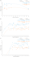
Regression models for the Log-number of bacteria (LNOB) recorded in external and internal tissues over time using T. dicentrarchi or T. maritimum specific qPCR assays. (A) is for the T. dicentrarchi treatment, (B) is for the T. maritimum high concentration treatment, (C) is for the T. maritimum low concentration treatment.
Figure 4
Figure 4
Histopathology of oral lesions from an Atlantic salmon smolt bath exposed to T. dicentrarchi. (A) Hematoxylin and eosin-stained sagittal section of the upper jaw with thick mats of thin filamentous bacteria (arrows) replacing areas of mucosa of the vomer and surrounding teeth. Black boxes in A and B indicate higher power magnification viewed in the next chronological alphabetical value. (B) Deep vomer ulceration overlying spongiotic epithelium (*) and necrotic submucosa (**). (C) Myriad long filamentous bacteria surrounding and replacing the submucosa around a tooth (t) of the vomer. (D) Oblique section of the lower jaw with fully exposed bone (*) with long filamentous bacterial mats (arrows) covering, replacing, and penetrating the oral mucosa and underlying viable and necrotic submucosa (**). Mats of similar bacteria (arrows) can be seen lining exposed bone and replacing dermal tissue on the underside of the jaw.
Figure 5
Figure 5
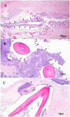
Histopathology of gill lesions from an Atlantic salmon smolt bath exposed to T. maritimum. (A) Hematoxylin and eosin-stained oblique section of the head and gills with necrotic and viable gill filaments covered by mats of long filamentous bacteria (arrows). Black boxes indicate higher power magnification as seen in (B,C). (B,C) Long filamentous bacteria (arrows) surrounding and penetrating blood vessels of a necrotic (*) and viable (**) gill filament, respectively.
Figure 6
Figure 6
Histopathology of an Atlantic salmon smolt bath exposed to T. maritimum. (A) Hematoxylin and eosin-stained oblique section of the caudal trunk of the fish with regionally extensive epidermal erosion, ulceration, and intralesional bacteria (arrows). Scale pocket edema is also present adjacent to the ulcer. Black boxes in A and B surround intralesional bacteria and outline the areas of higher magnification as seen in (B,C). (B) Epidermis overlying scales is absent and bacterial mats (arrows) can be seen under scales. (C) Filamentous bacteria (arrows) replacing submucosa between scales and complete loss of epithelium on the external scale surface. (D) Large, deep epidermal ulceration of the lateral trunk of the fish with thick mats of intralesional bacteria (arrows) covering and penetrating (*) exposed dermis and underlying musculature.
Figure 7
Figure 7
Histopathology of an Atlantic salmon smolt bath exposed to T. maritimum. (A) Hematoxylin and eosin-stained sagittal section of the buccopharyngeal cavity with extensive replacement of oral mucosa by thick mats of long filamentous bacteria (arrows). Sections of the submucosa (s) and deep muscle (m) bundles are undergoing degeneration and necrosis. Black boxes indicate higher power magnification as seen in (B,C). (B,C) Long filamentous bacteria (arrows) penetrating necrotic submucosa and surrounding buccopharyngeal teeth (t).

References

    1. Nowlan J.P., Lumsden J.S., Russell S. Advancements in characterizing Tenacibaculum infections in Canada. Pathogens. 2020;9:1029. doi: 10.3390/pathogens9121029. - DOI - PMC - PubMed
    1. Avendaño-Herrera R., Toranzo A.E., Magariños B. Tenacibaculosis infection in marine fish caused by Tenacibaculum maritmum: A review. Dis. Aquat. Org. 2006;71:255–266. doi: 10.3354/dao071255. - DOI - PubMed
    1. Avendaño-Herrera R., Collarte C., Saldarriaga-Córdoba M., Irgang R. New salmonid hosts for Tenacibaculum species: Expansion of tenacibaculosis in Chilean aquaculture. J. Fish Dis. 2020;43:1077–1108. doi: 10.1111/jfd.13213. - DOI - PubMed
    1. Avendaño-Herrera R., Irgang R., Sandoval C., Moreno-Lira P., Houel A., Duchaud E., Ilardi P. Isolation, characterization and virulence potential of Tenacibaculum dicentrarchi in salmonid cultures in Chile. Transbound. Emerg. Dis. 2016;63:121–126. doi: 10.1111/tbed.12464. - DOI - PubMed
    1. Frisch K., Småge S.B., Johansen R., Duesund H., Brevik Ø.J., Nylund A. Pathology of experimentally induced mouthrot caused by Tenacibaculum maritimum in Atlantic salmon smolts. PLoS ONE. 2018;13:e0206951. doi: 10.1371/journal.pone.0206951. - DOI - PMC - PubMed

LinkOut - more resources