Review
. 2021 Nov 6;10(11):1447.
doi: 10.3390/pathogens10111447.

The Global Emergence of Human Babesiosis

Affiliations
Review

The Global Emergence of Human Babesiosis

Abhinav Kumar et al. Pathogens. .

Erratum in

Abstract

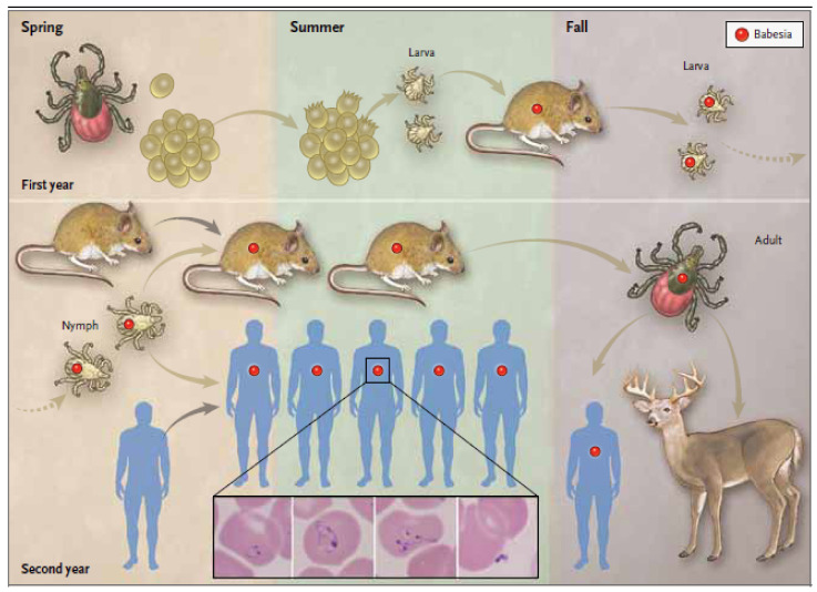
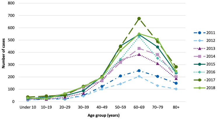
Babesiosis is an emerging tick-borne disease caused by intraerythrocytic protozoa that are primarily transmitted by hard-bodied (ixodid) ticks and rarely through blood transfusion, perinatally, and organ transplantation. More than 100 Babesia species infect a wide spectrum of wild and domestic animals worldwide and six have been identified as human pathogens. Babesia microti is the predominant species that infects humans, is found throughout the world, and causes endemic disease in the United States and China. Babesia venatorum and Babesia crassa-like agent also cause endemic disease in China. Babesia divergens is the predominant species in Europe where fulminant cases have been reported sporadically. The number of B. microti infections has been increasing globally in recent decades. In the United States, more than 2000 cases are reported each year, although the actual number is thought to be much higher. In this review of the epidemiology of human babesiosis, we discuss epidemiologic tools used to monitor disease location and frequency; demographics and modes of transmission; the location of human babesiosis; the causative Babesia species in the Americas, Europe, Asia, Africa, and Australia; the primary clinical characteristics associated with each of these infections; and the increasing global health burden of this disease.

Keywords: Babesia microti; babesiosis; case surveillance; epidemiology; immunoepidemiology.


Conflict of interest statement

The authors declare no conflict of interest.

Figures

Figure 1
Figure 1
Geographic distribution of major areas of human babesiosis transmission. The map depicts the major areas where human babesiosis has been reported. Additional areas where human babesiosis has been reported but are not shown in the figure are mentioned in the text. Solid colors indicate areas where human babesiosis is endemic. Stippled areas indicate areas where babesiosis is sporadic with ≥10 cases reported. Circles depict areas where 1–10 cases have been reported. Colors distinguish the etiologic agents: Babesia crassa-like agent (gray), Babesia duncani (orange), Babesia divergens (blue), Babesia microti (red), Babesia motasi (black), and Babesia venatorum (green). White circles depict cases caused by Babesia spp. that were not characterized. Asymptomatic infections are omitted (adapted from The New England Journal of Medicine, Edouard Vannier, and Peter J. Krause, Human Babesiosis, 2012, 366, 2397. Copyright (2021) Massachusetts Medical Society. Reprinted with permission [1]).
Figure 2
Figure 2
Transmission of Babesia microti and stages in the Ixodes scapularis tick vector life cycle. Female I. scapularis lay 2000–3000 eggs in the spring that hatch in early summer and produce larvae. Larval I. scapularis ticks become infected with B. microti when they take a blood meal from infected white-footed mice (Peromyscus leucopus) or other small rodent hosts in late summer. Fed larvae molt into nymphs and overwinter. During the following late spring, summer, and early autumn, infected nymphs transmit B. microti to uninfected mice or humans when they take a blood meal. In the autumn, nymphs molt into adults. Adult males and females preferentially feed and procreate on white-tailed deer (Odocoileus virginianus) but rarely on humans. The blood meal provides sufficient protein for female ticks to lay eggs. The tick life cycle is repeated when a new generation of larvae hatch from the eggs in the early spring to complete the tick life cycle. Deer do not become infected with B. microti. The inset panels from left to right show a B. microti ring form with a non-staining vacuole surrounded by cytoplasm (blue) and two small nuclei (purple), an amoeboid form, a tetrad form (also referred to as a Maltese cross), and an extracellular form (adapted from The New England Journal of Medicine, Edouard Vannier, and Peter J. Krause, Human Babesiosis, 2012, 366, 2397. Copyright (2021) Massachusetts Medical Society. Reprinted with permission [1]).
Figure 3
Figure 3
Babesiosis cases by age in the United States. Babesiosis cases reported by age to the Centers for Disease Control and Prevention, United States between 2011 and 2018 are shown. The low numbers of cases in children is due to the mild clinical symptoms resulting from B. microti infection rather than exposure to the infection. Almost half of children are asymptomatically infected compared to about a fifth of adults. Thus, B. microti-infected children are not diagnosed as frequently as adults (adapted from the Centers for Disease Control and Prevention. Notifiable Diseases and Mortality Tables. MMWR Morb Mortal Wkly Rep 2016, 65(3) [95]).
Figure 4
Figure 4
Human babesiosis occurs within Lyme disease endemic areas in the United States. Lyme disease and human babesiosis have been nationally notifiable conditions since 1991 and 2011, respectively. The names of counties that reported cases of Lyme disease and/or babesiosis from 2011 to 2013 were obtained from the Centers for Disease Control and Prevention. Counties with three or more cases of Lyme disease but fewer than three cases of babesiosis are depicted in green. Counties with three or more cases of Lyme disease and three or more cases of babesiosis are depicted in gray. No county reported three or more cases of babesiosis but fewer than three cases of Lyme disease (adapted from Diuk-Wasser M, Vannier E, Krause PJ. Coinfection by Ixodes tick-borne pathogens: Ecological, epidemiological, and clinical consequences. Trends Parasitol 2016, 32, 30–42 [43]).

References

    1. Vannier E., Krause P.J. Human babesiosis. N. Engl. J. Med. 2012;366:2397–2407. doi: 10.1056/NEJMra1202018. - DOI - PubMed
    1. Hildebrandt A., Zintl A., Montero E., Hunfeld K.P., Gray J. Human babesiosis in Europe. Pathogens. 2021;10:1165. doi: 10.3390/pathogens10091165. - DOI - PMC - PubMed
    1. Fang L.Q., Liu K., Li X.L., Liang S., Yang Y., Yao H.W., Sun R.X., Sun Y., Chen W.J., Zuo S.Q., et al. Emerging tick-borne infections in mainland China: An increasing public health threat. Lancet Infect. Dis. 2015;15:1467–1479. doi: 10.1016/S1473-3099(15)00177-2. - DOI - PMC - PubMed
    1. Krause P.J. Human babesiosis. Int. J. Parasitol. 2019;49:165–174. doi: 10.1016/j.ijpara.2018.11.007. - DOI - PubMed
    1. Hunfeld K.P., Hildebrandt A., Gray J.S. Babesiosis: Recent insights into an ancient disease. Int. J. Parasitol. 2008;38:1219–1237. doi: 10.1016/j.ijpara.2008.03.001. - DOI - PubMed

LinkOut - more resources