Review
. 2021 Oct 21;11(11):1119.
doi: 10.3390/life11111119.

Metabolism in Retinopathy of Prematurity

Affiliations
Review

Metabolism in Retinopathy of Prematurity

Yohei Tomita et al. Life (Basel). .

Abstract

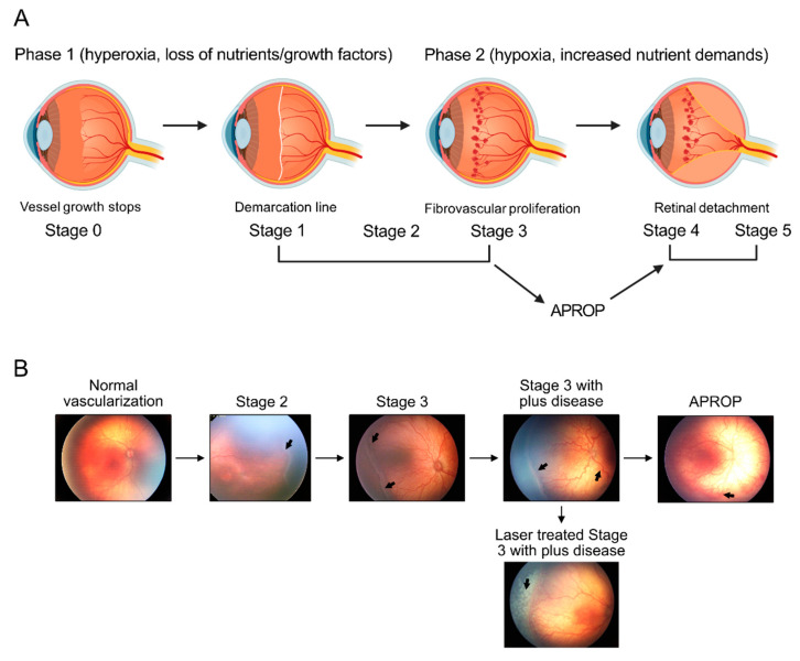
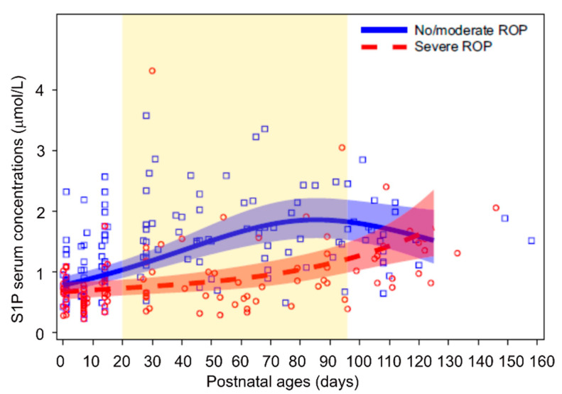
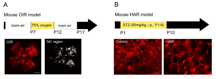
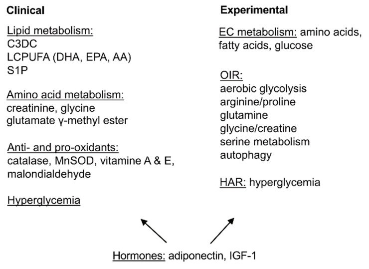
Retinopathy of prematurity is defined as retinal abnormalities that occur during development as a consequence of disturbed oxygen conditions and nutrient supply after preterm birth. Both neuronal maturation and retinal vascularization are impaired, leading to the compensatory but uncontrolled retinal neovessel growth. Current therapeutic interventions target the hypoxia-induced neovessels but negatively impact retinal neurons and normal vessels. Emerging evidence suggests that metabolic disturbance is a significant and underexplored risk factor in the disease pathogenesis. Hyperglycemia and dyslipidemia correlate with the retinal neurovascular dysfunction in infants born prematurely. Nutritional and hormonal supplementation relieve metabolic stress and improve retinal maturation. Here we focus on the mechanisms through which metabolism is involved in preterm-birth-related retinal disorder from clinical and experimental investigations. We will review and discuss potential therapeutic targets through the restoration of metabolic responses to prevent disease development and progression.

Keywords: dyslipidemia; hyperglycemia; hyperglycemia-associated retinopathy; neovascularization; oxygen-induced retinopathy; retinal metabolism; retinopathy of prematurity.


Conflict of interest statement

The authors declare no conflict of interest.

Figures

Figure 1
Figure 1
ROP progression in premature infants. (A) Schematics of the progression of human retinopathy of prematurity (ROP). Phases 1 and 2 of ROP are associated with different oxygen levels. Loss of essential nutrients and pro-angiogenic growth factors after birth in combination with provision of high supplemental oxygen, leads to hyperoxia that suppresses retinal vascularization (Phase 1). In the second phase of ROP (Phase 2), relative hypoxia and increased nutrient demands of the avascular retina drives fibrovascular proliferation. ROP Phase 2 is defined by anatomic changes, such as the demarcation line (stage 1), ridge (stage 2), extraretinal fibrovascular proliferation (stage 3), partial retinal detachment (stage 4), and total retinal detachment (stage 5). Any stage can develop into aggressive posterior ROP (APROP), which rapidly progresses to tractional retinal detachment (stage 4 or 5). Image made with graphics from ©BioRender (https://biorender.com/ (accessed on 18 October 2021) Agreement number: IA22XF3W0H) (B) Illustration of retinopathy of prematurity (ROP) development, from normal retinal neuro-vascular development, via stage 2 with ridge (arrow), stage 3 with neovascularization and hemorrhage (arrows), stage 3 with plus disease (arrow), APROP with central changes (arrow) and laser treatment (arrow) of stage 3 ROP.
Figure 2
Figure 2
Serum S1P levels and ROP in premature infants. Dots show measured serum S1P levels and lines (with 95% CI) represent estimates from mixed model for repeated measures adjusted for GA at birth and weight standard deviation score. Graph area highlighted in yellow represents time points where curves differ significantly after adjustment for multiplicity. n = 28 for no/moderate ROP, n = 19 for severe ROP. Graph was adapted from [42].
Figure 3
Figure 3
Mouse models of ROP. (A) Mouse model of oxygen-induced retinopathy (OIR). Mouse neonates at postnatal day (P) 7 with their nursing dam are exposed to 75% oxygen for five days. At P12, mice are returned to room air. At P17, neovascularization (NV) reaches the maximum. Retinal vasculature is visualized with isolectin (red) staining and NV area is highlighted in white. Scale bar, 1 mm. (B) Mouse model of hyperglycemia-associated retinopathy (HAR). Hyperglycemia is induced in mouse neonates with streptozotocin (STZ, 50 mg/kg) intraperitoneally (i.p.) from P1 to P9. At P10, retinal vessels in the deep vascular plexus are visualized with isolectin (red) staining. Reduced retinal vascular density is observed in HAR. Scale bar, 50 µm.
Figure 4
Figure 4
Summarized current findings of metabolism in ROP. Lipid and amino acid metabolic disturbance, hyperglycemia as well as unbalanced antioxidant system were found. Hormones including adiponectin and IGF-1 are essential in modulating metabolic responses. The combination of nutrients and the timing of nutritional and hormonal intervention, as well as the corresponding specific cell responses need to be carefully evaluated.

References

    1. Hellstrom A., Smith L.E., Dammann O. Retinopathy of prematurity. Lancet. 2013;382:1445–1457. doi: 10.1016/S0140-6736(13)60178-6. - DOI - PMC - PubMed
    1. Fu Z., Gong Y., Lofqvist C., Hellstrom A., Smith L.E. Review: Adiponectin in retinopathy. Biochim. Biophys. Acta. 2016;1862:1392–1400. doi: 10.1016/j.bbadis.2016.05.002. - DOI - PMC - PubMed
    1. Joyal J.S., Sun Y., Gantner M.L., Shao Z., Evans L.P., Saba N., Fredrick T., Burnim S., Kim J.S., Patel G., et al. Retinal lipid and glucose metabolism dictates angiogenesis through the lipid sensor Ffar1. Nat. Med. 2016;22:439–445. doi: 10.1038/nm.4059. - DOI - PMC - PubMed
    1. Smith L.E., Wesolowski E., McLellan A., Kostyk S.K., D’Amato R., Sullivan R., D’Amore P.A. Oxygen-induced retinopathy in the mouse. Investig. Ophthalmol. Vis. Sci. 1994;35:101–111. - PubMed
    1. Hansen R.M., Moskowitz A., Akula J.D., Fulton A.B. The neural retina in retinopathy of prematurity. Prog. Retin. Eye Res. 2017;56:32–57. doi: 10.1016/j.preteyeres.2016.09.004. - DOI - PMC - PubMed

LinkOut - more resources