Case Reports
. 2021 Nov 9;57(11):1220.
doi: 10.3390/medicina57111220.

Limb Salvage after Lower-Leg Fracture and Popliteal Artery Transection-The Role of Vessel-First Strategy and Bone Fixation Using the Ilizarov External Fixator Device: A Case Report

Affiliations
Case Reports

Limb Salvage after Lower-Leg Fracture and Popliteal Artery Transection-The Role of Vessel-First Strategy and Bone Fixation Using the Ilizarov External Fixator Device: A Case Report

Vincenzo Giordano et al. Medicina (Kaunas). .

Abstract

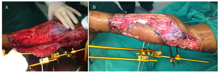
Open traumatic lesion of the popliteal artery is relatively rare. Ischemia time longer than 6 h and severity of limb ischemia have been shown to be associated with an increased risk of limb loss. Severe local infection is critical in the presence of major soft tissue trauma or open fractures. We report the case of a young female who suffered a traumatic transection of the popliteal artery associated with an open fracture of the distal tibia and fibula managed by direct vessel reconstruction with an end-to-end repair and skeletal stabilization initially with half-pin external fixation, then replaced by an Ilizarov circular frame. The patient had a very satisfactory outcome, but the fracture healed malunited, later corrected by open reduction and internal fixation with lag-screwing and a neutralization plate.

Keywords: Ilizarov circular fixator; external fixation; limb ischemia; malunion; popliteal artery.


Conflict of interest statement

The authors declare no conflict of interest related to the report of the case.

Figures

Figure 1
Figure 1
(A) Severe degloving of the left leg. (B) Lateral radiograph of the left leg, showing an open simple transverse distal tibia and fibula fracture.
Figure 2
Figure 2
(A) The open wound was initially debrided, and a half-pin spanning external fixator was applied on the anterior aspect of the left lower limb, with the knee in slight flexion (approximately 10°) and the ankle in neutral position. (B) After identification of the complete transection in the middle part of the popliteal artery, vascular surgeons performed direct vessel reconstruction with an end-to-end repair, and the skin flaps were loosely approximated and sutured to the gastrocnemius muscle.
Figure 3
Figure 3
(A,B) Anterior and medial aspects of the left leg on day 2, demonstrating a large area of necrosis in the antero-medial part of the left leg. (C) The skin flap used to cover the degloved area of the leg was completely removed, and the wound was extensively debrided and irrigated.
Figure 4
Figure 4
(A) Aspect of the leg on day 4. (B) Aspect of the leg on day 12. (C) Aspect of the leg on day 30, when half-pin external fixator was removed and an Ilizarov circular frame was applied to hold the distal tibia in place until bone healing.
Figure 5
Figure 5
Aspect of the leg one week after the Ilizarov circular frame was applied. No wound closure either using NPWT, flaps, or skin graft was done. There were no signs of ischemia or vascular insufficiency.
Figure 6
Figure 6
AP and lateral radiographic views of the left leg demonstrating the adequate alignment of the fracture and slight progression of bone healing, with minimum varus on the fracture site.
Figure 7
Figure 7
On day 100, the fracture and the leg wound were totally healed, and the patient was admitted for removal of the Ilizarov frame. Radiographs showed malunited fractures of the left distal tibia and fibula.
Figure 8
Figure 8
Immediate postoperative radiographic images of the one-stage corrective osteotomy of the malunited fractures. A 2 cm resection of the distal fibula was initially performed, followed by an oblique osteotomy of the distal tibia through an antero-medial approach.
Figure 9
Figure 9
Radiographs taken in the last follow-up revealed a satisfactory alignment of the left leg on both planes.

References

    1. Fairhurst P.G., Wyss T.R., Weiss S., Becker D., Schmidli J., Makaloski V. Popliteal Vessel Trauma: Surgical Approaches and the Vessel-First Strategy. Knee. 2018;25:849–855. doi: 10.1016/j.knee.2018.06.012. - DOI - PubMed
    1. O’Banion L.A., Dirks R., Saldana-Ruiz N., Farooqui E., Yoon W.J., Pozolo C., Fox C.J., Crally A., Siada S., Nehler M.R., et al. Contemporary Outcomes of Traumatic Popliteal Artery Injury Repair from the Popliteal Scoring Assessment for Vascular Extremity Injury in Trauma Study. J. Vasc. Surg. 2021;74:1573–1580.e2. doi: 10.1016/j.jvs.2021.04.064. - DOI - PubMed
    1. Perkins Z.B., Yet B., Glasgow S., Cole E., Marsh W., Brohi K., Rasmussen T., Tai N.R.M. Meta-Analysis of Prognostic Factors for Amputation Following Surgical Repair of Lower Extremity Vascular Trauma. BJS. 2015;102:436–450. doi: 10.1002/bjs.9689. - DOI - PubMed
    1. Liu Y.-W., Li Y.-H., Yu T., Yang T., Li Y., Tan L. Popliteal Artery Transection Associated with a Minimally Displaced Tibial Plateau Fracture: A Case Report and Review of the Literature. BMC Musculoskelet. Disord. 2020;21:59. doi: 10.1186/s12891-020-3089-8. - DOI - PMC - PubMed
    1. Bliven E.K., Greinwald M., Hackl S., Augat P. External Fixation of the Lower Extremities: Biomechanical Perspective and Recent Innovations. Injury. 2019;50:S10–S17. doi: 10.1016/j.injury.2019.03.041. - DOI - PubMed

Publication types