. 2021 Nov 14;26(22):6863.
doi: 10.3390/molecules26226863.

Virtual Screening for Potential Phytobioactives as Therapeutic Leads to Inhibit NQO1 for Selective Anticancer Therapy

Affiliations

Virtual Screening for Potential Phytobioactives as Therapeutic Leads to Inhibit NQO1 for Selective Anticancer Therapy

Bhargav Shreevatsa et al. Molecules. .

Abstract

NAD(P)H:quinone acceptor oxidoreductase-1 (NQO1) is a ubiquitous flavin adenine dinucleotide-dependent flavoprotein that promotes obligatory two-electron reductions of quinones, quinonimines, nitroaromatics, and azo dyes. NQO1 is a multifunctional antioxidant enzyme whose expression and deletion are linked to reduced and increased oxidative stress susceptibilities. NQO1 acts as both a tumor suppressor and tumor promoter; thus, the inhibition of NQO1 results in less tumor burden. In addition, the high expression of NQO1 is associated with a shorter survival time of cancer patients. Inhibiting NQO1 also enables certain anticancer agents to evade the detoxification process. In this study, a series of phytobioactives were screened based on their chemical classes such as coumarins, flavonoids, and triterpenoids for their action on NQO1. The in silico evaluations were conducted using PyRx virtual screening tools, where the flavone compound, Orientin showed a better binding affinity score of -8.18 when compared with standard inhibitor Dicumarol with favorable ADME properties. An MD simulation study found that the Orientin binding to NQO1 away from the substrate-binding site induces a potential conformational change in the substrate-binding site, thereby inhibiting substrate accessibility towards the FAD-binding domain. Furthermore, with this computational approach we are offering a scope for validation of the new therapeutic components for their in vitro and in vivo efficacy against NQO1.

Keywords: NQO1; Nrf2; binding affinity; conceptual DFT; detoxification; molecular docking; phytobiactives; virtual screening.


Conflict of interest statement

The authors declare no conflict of interest.

Figures

Figure 1
Figure 1
Tumor suppressor and tumor promoter behavior of NQO1 linked to reduced and increased oxidative stress susceptibilities.
Figure 2
Figure 2
In some cases, (A) the expression levels of NQO1 (yellow circles) in a normal cell play a vital role in protecting the cells against harmful components; (B) in cancer cells, the over expression of NQO1 detoxifies Quinine molecules (green circles) resulting as Quinone resistant; (C) the inhibition of NQO1 in cancer cells avoids Quinone detoxification and shows susceptibility to anti-cancerous agents.
Figure 3
Figure 3
Diagrammatic representation of therapeutic approach with reduction of NQO1 levels in tumor cells results in decreased tumor burden: (A) normal liver expressing NQO1 (yellow circles) at normal level; (B) NQO1 (yellow circles) over expressed in tumor cells; (C) treating with anti-NQO1 drugs leads to the decreased tumor burden by decreased level of NQO1 (inhibited NQO1 black circles).
Figure 4
Figure 4
Two-dimensional and three-dimensional structures of major phytobioactives.
Figure 5
Figure 5
Three-dimensional structure of the protein NQO1 (PDB ID: 2F1O).
Figure 6
Figure 6
The Ramachandran plot generated from RAMPAGE. The Ramachandran plot representing energetically allowable regions for backbone dihedral angles ψ vs. ϕ amino acid residues in NQO1 (2F1O) protein structure.
Figure 7
Figure 7
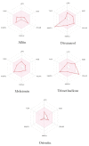
Bioavailabity radars of phytobioactives based on physicochemical indices with ideal values for oral bioavailability.
Figure 8
Figure 8
Histogram showing the Molecular Docking results between NQO1 (2F1O) against selective phytobioactives (the binding energy value δG is shown in minus kcal/mol).
Figure 9
Figure 9
A Comparative study of the standard NQO1 inhibitor Dicumarol with the other potent phytobioactives. The X-Axis: Phytobioactives v/s Y-Axis: Number of hydrogen interactions towards protein.
Figure 10
Figure 10
The putative binding pose of Orientin with NQO1: (a) Orientin represented in green can be seen deeply embedded into the active site. (b) The two-dimensional geometrical pose of Orientin interacting with surrounding amino acids at the putative binding cavity of NQO1.
Figure 11
Figure 11
2-D and 3-D structures of several phytobioactives docked with NQO1.
Figure 12
Figure 12
Graphical representation of (a) root-mean-square fluctuation for Orientin with active site residues of NQO1 after MDS and (b) ligand RMSD plot to show the contact point pf the ligand with the amino residue of the NQO1 represented as green color vertical green lines with several contact points.
Figure 13
Figure 13
A scheme of detailed Orientin atom interactions with the protein residues. (a) Interactions that occur more than 30% of the simulation time in the selected trajectory. (b) Normalized stacked bar chart of compound Orientin interacting with NQO1 away from active site pocket through a hydrogen bond, hydrophobic and ionic interactions, and water bridges, and (c) The number of contact points with the amino acid residues are depicted in timeline representations coded with color intensity graphs throughout the simulation.
Figure 14
Figure 14
Protein secondary structure elements (SSE) like α-helices and β-strands are monitored throughout the simulation. The plot above reports SSE distribution by residue index throughout the protein structure.
Figure 15
Figure 15
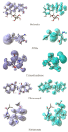
Graphical Representations of the Dual Descriptor DD of the Five Studied Ligands. (Left): DD > 0, (Right): DD < 0.

References

    1. Dinkova-Kostova A.T., Talalay P. NAD(P)H:quinone Acceptor Oxidoreductase 1 (NQO1), a Multifunctional Antioxidant Enzyme and Exceptionally Versatile Cytoprotector. Arch. Biochem. Biophys. 2010;501:116–123. doi: 10.1016/j.abb.2010.03.019. - DOI - PMC - PubMed
    1. Ross D., Siegel D. Functions of NQO1 in Cellular Protection and CoQ10 Metabolism and its Potential Role as a Redox Sensitive Molecular Switch. Front. Physiol. 2017;8:595. doi: 10.3389/fphys.2017.00595. - DOI - PMC - PubMed
    1. Oh E.T., Park H.J. Implications of NQO1 in Cancer Therapy. BMB Rep. 2015;48:609–617. doi: 10.5483/BMBRep.2015.48.11.190. - DOI - PMC - PubMed
    1. Asher G., Dym O., Tsvetkov P., Adler J., Shaul Y. The Crystal Structure of NAD(P)H Quinone Oxidoreductase 1 in Complex with Its Potent Inhibitor Dicoumarol. Biochemistry. 2006;45:6372–6378. doi: 10.1021/bi0600087. - DOI - PubMed
    1. Siegel D., Ross D. Immunodetection of NAD(P)H:quinone Oxidoreductase 1 (NQO1) in Human Tissues. Free Radic. Biol. Med. 2000;29:246–253. doi: 10.1016/S0891-5849(00)00310-5. - DOI - PubMed

MeSH terms