Review
. 2021 Nov 15;26(22):6876.
doi: 10.3390/molecules26226876.

Malate Transport and Metabolism in Nitrogen-Fixing Legume Nodules

Affiliations
Review

Malate Transport and Metabolism in Nitrogen-Fixing Legume Nodules

Nicholas J Booth et al. Molecules. .

Abstract

Legumes form a symbiosis with rhizobia, a soil bacterium that allows them to access atmospheric nitrogen and deliver it to the plant for growth. Biological nitrogen fixation occurs in specialized organs, termed nodules, that develop on the legume root system and house nitrogen-fixing rhizobial bacteroids in organelle-like structures termed symbiosomes. The process is highly energetic and there is a large demand for carbon by the bacteroids. This carbon is supplied to the nodule as sucrose, which is broken down in nodule cells to organic acids, principally malate, that can then be assimilated by bacteroids. Sucrose may move through apoplastic and/or symplastic routes to the uninfected cells of the nodule or be directly metabolised at the site of import within the vascular parenchyma cells. Malate must be transported to the infected cells and then across the symbiosome membrane, where it is taken up by bacteroids through a well-characterized dct system. The dicarboxylate transporters on the infected cell and symbiosome membranes have been functionally characterized but remain unidentified. Proteomic and transcriptomic studies have revealed numerous candidates, but more work is required to characterize their function and localise the proteins in planta. GABA, which is present at high concentrations in nodules, may play a regulatory role, but this remains to be explored.

Keywords: legume; malate; metabolism; nitrogen fixation; nodules.


Conflict of interest statement

The authors declare no conflict of interest.

Figures

Figure 1
Figure 1
Structure of determinate (a) and indeterminate (b) nodules. a. Section of a determinate nodule from soybean. Determinate nodules do not have a persistent meristem. The central infected zone contains large infected cells (I) containing symbiosomes and smaller uninfected cells (U). This region is surrounded by the cortex including the inner cortex (IC) with vascular bundles (VB) and the outer cortex (OC). (b). Section of an indeterminate M. truncatula nodule. The nodule has a persistent meristem (M, often termed zone I) and continues to grow producing zones of different developmental stages including the infection zone (IZ, zone II), transition zone (TZ, interzone II-III), nitrogen fixation zone (FZ, zone IV) and senescence zone (SZ, zone V). The nitrogen fixation zone contains both infected cells (I) and uninfected cells. Vascular bundles (VB) are present in the nodule cortex (C). Images courtesy of Aleksandr Gavrin.
Figure 2
Figure 2
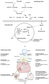
Nodule carbon metabolism. (a) The metabolism of sucrose to produce malate is likely to be in uninfected cells but may also occur in infected cells. In infected cells malate is transported to the symbiosome to support nitrogen fixation by bacteroids or to mitochondria, where it is used to generate ATP and the carbon skeletons required for nitrogen assimilation. Arrows with dashed lines indicate reactions that may occur but are unlikely to be significant in production of malate. PEP: phosphoenolpyruvate; PEPC: PEP carboxylase; OAA: oxaloacetate; TCAC: tricarboxylate cycle; mETC: mitochondrial electron transport chain. (b) Summary of gene expression, enzyme and transporter localisation in nodules and putative pathways for sucrose and malate movement within the nodule. Apoplastic routes are suggested by the presence of LjALMT4, LjSWEET3 and MtSWEET11 on the plasma membrane of nodule vascular parenchyma [24,25,26]. Malate transporters have been characterized on the infected cell plasma membrane [27] and symbiosome membrane [28] but the proteins encoding them have not been described. Malate is transported into bacteroids by a dicarboxylate transporter DctA, that is upregulated under symbiotic conditions [29,30]. Malate must be imported into the mitochondria but the transporter has not been described [31,32,33,34]. A sucrose importer may be present on infected and/or uninfected cell plasma membranes to support the activity of SS [35]. Symplastic routes are suggested by presence of plasmodesmata between cells in the nodule and studies with microinjection of Lucifer Yellow-CH and trafficking studies with GFP [20,21,22]. Ps: Pisum sativum, Mt: M. truncatula, Rl: Rhizobium leguminosarum, Lj: L. japonicus, Gm: G. max, Pv: Phaseolus vulgaris, Vu: Vigna unguiculata, tpr: transporter. Short dashed lines indicate possible pathways for malate movement. Long dashed lines indicate possible pathways for sucrose movement. Figure created with BioRender.com (accessed on 1 November 2021).

Similar articles

Cited by

References

    1. Oldroyd G.E., Murray J.D., Poole P.S., Downie J.A. The rules of engagement in the legume-rhizobial symbiosis. Annu. Rev. Gen. 2011;45:119–144. doi: 10.1146/annurev-genet-110410-132549. - DOI - PubMed
    1. Whitehead L.F., Day D.A. The peribacteroid membrane. Physiol. Plant. 1997;100:30–44. doi: 10.1111/j.1399-3054.1997.tb03452.x. - DOI
    1. Udvardi M., Poole P.S. Transport and Metabolism in Legume-Rhizobia Symbioses. Annu. Rev. Plant Biol. 2013;64:781–805. doi: 10.1146/annurev-arplant-050312-120235. - DOI - PubMed
    1. Gavrin A., Kaiser B.N., Geiger D., Tyerman S.D., Wen Z., Bisseling T., Fedorova E.E. Adjustment of Host Cells for Accommodation of Symbiotic Bacteria: Vacuole Defunctionalization, HOPS Suppression, and TIP1g Retargeting in Medicago. Plant Cell. 2014;26:3809. doi: 10.1105/tpc.114.128736. - DOI - PMC - PubMed
    1. Mohd Noor S., Day D., Smith P. The Symbiosome Membrane. In: Bruijn F.J., editor. Biological Nitrogen Fixation. John Wiley & Sons; Hoboken, NJ, USA: 2015. pp. 683–694.

LinkOut - more resources