. 2021 Oct 21;13(11):1754.
doi: 10.3390/pharmaceutics13111754.

Green Synthesized Silver Nanoparticles Using Tridax Procumbens for Topical Application: Excision Wound Model and Histopathological Studies

Affiliations

Green Synthesized Silver Nanoparticles Using Tridax Procumbens for Topical Application: Excision Wound Model and Histopathological Studies

Farhat Fatima et al. Pharmaceutics. .

Abstract

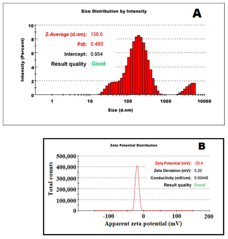
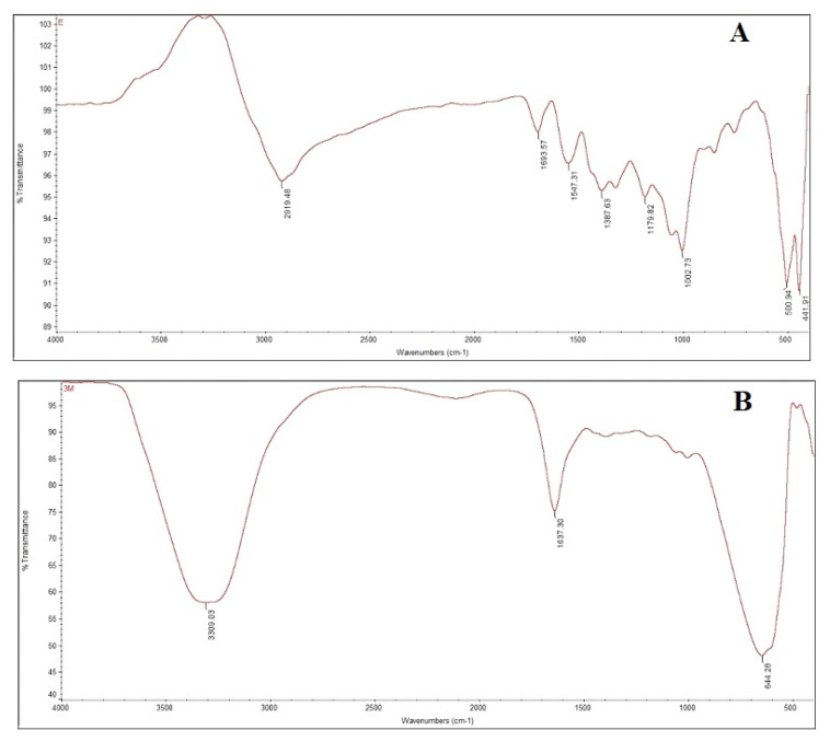
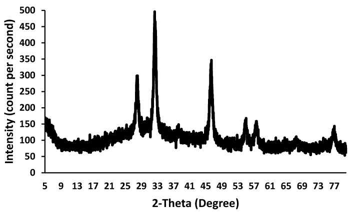
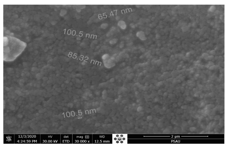
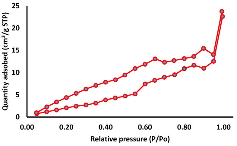
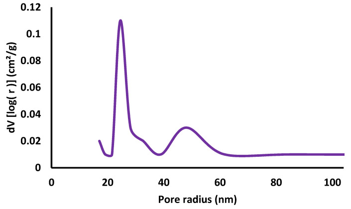
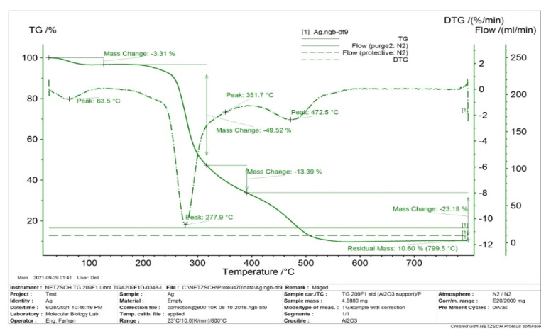
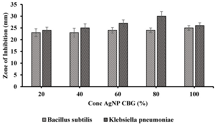
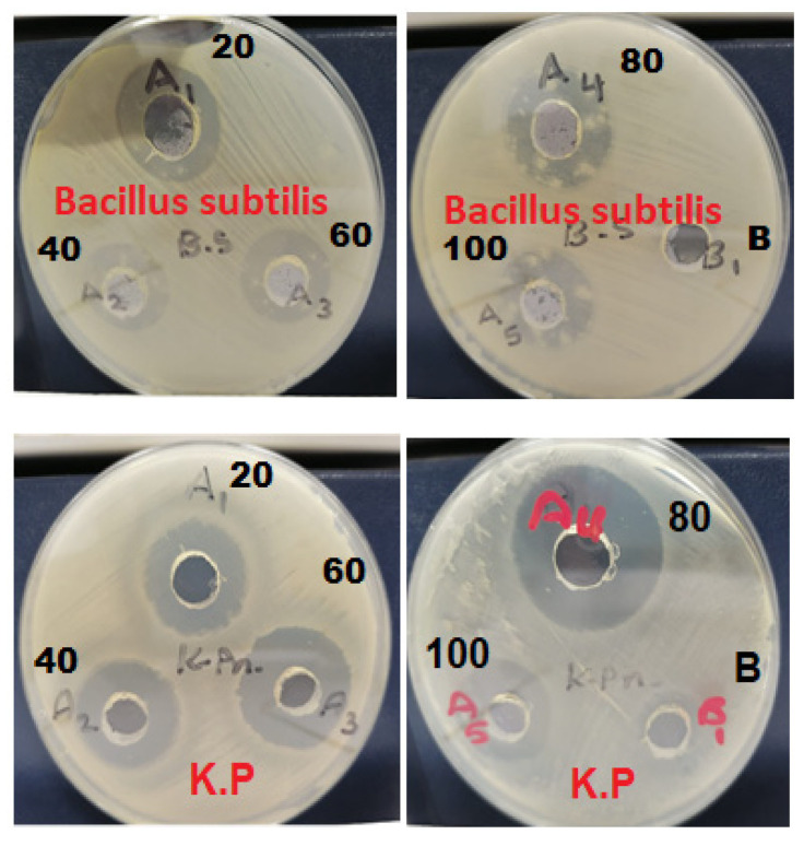
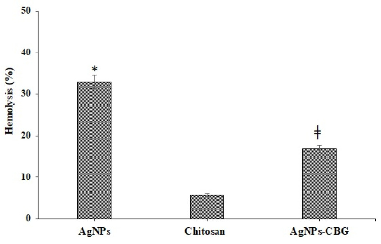
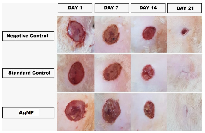
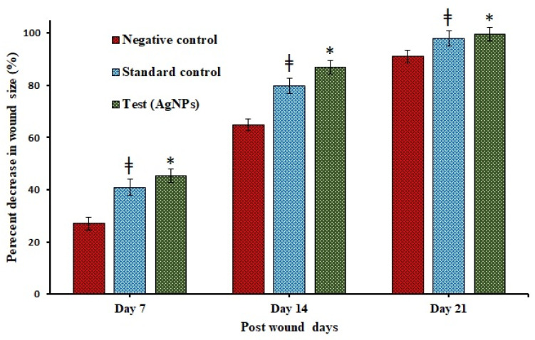
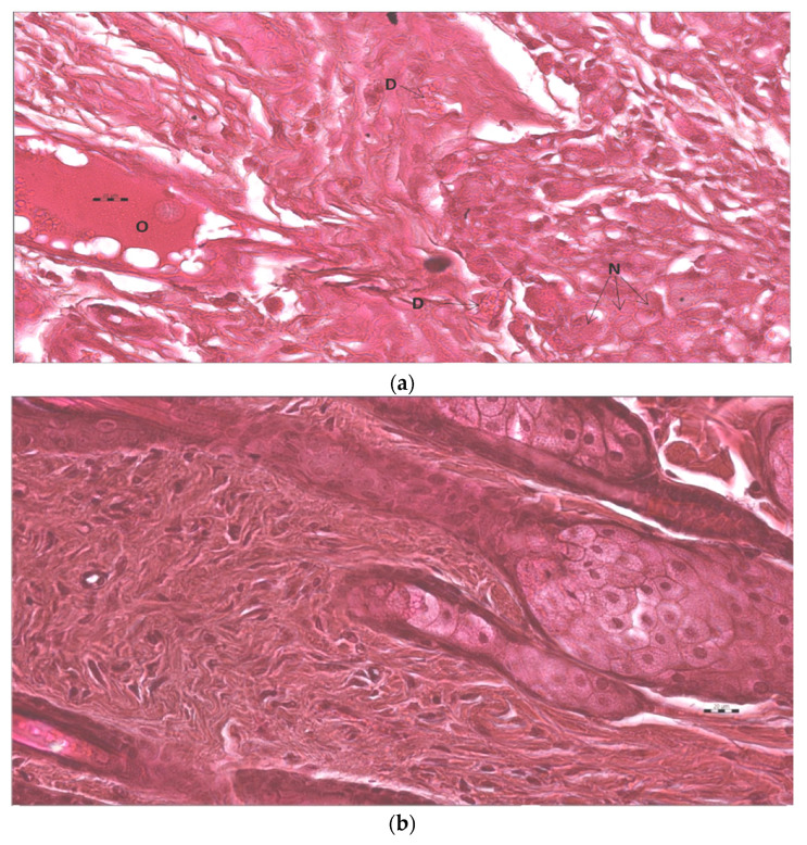
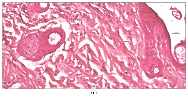
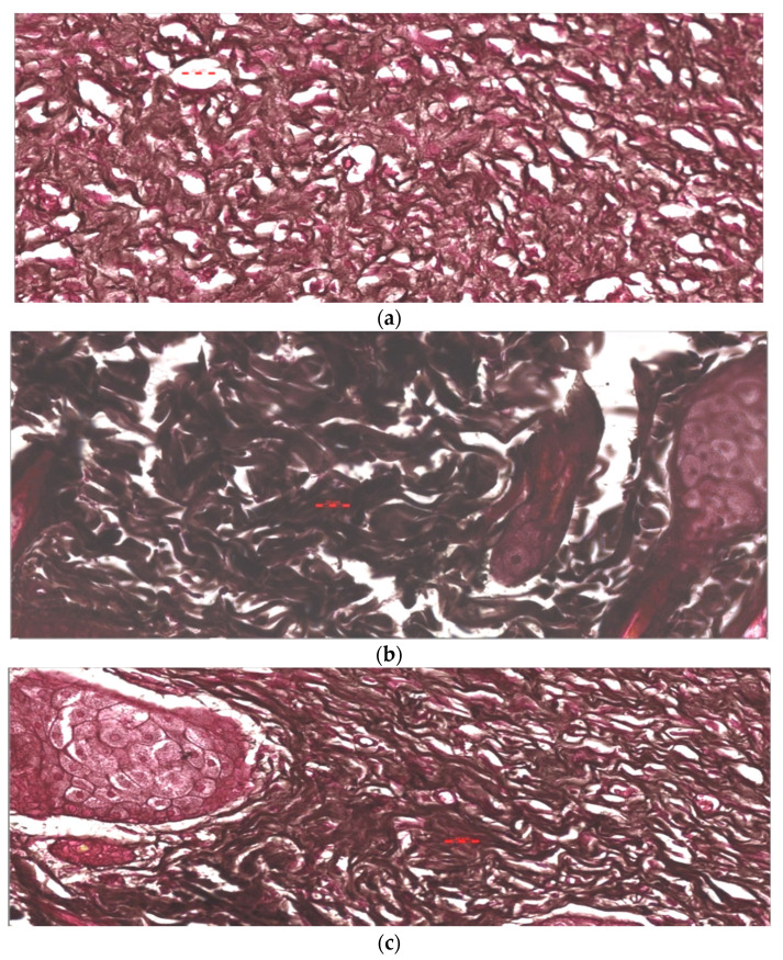
The objective of this study was to synthesize silver nanoparticles from the leaves of Tridax procumbens and develop its topical gels using chitosan to investigate the wound healing efficacy concomitant with the histopathological study. Green synthesized silver nanoparticles (AgNPs) were prepared by reacting silver nitrate (0.3 M) with leaf extract and characterized by particle analysis, FTIR, XRD, SEM, BET, and TGA. The results revealed formed AgNPs were nano-sized (138 ± 2.1 nm), monodispersed (PDI: 0.460 ± 0.3), inter-particle repulsion (zeta: -20.4 ± 5.20 mV), stabilized, crystalline and, spherical with size ranging from 80-100 nm as per SEM micro photos. The BET analysis of AgNPs presents the surface area (12.861 m2/g), pore volume (0.037 cc/g), and pore radius (24.50 nm).TGA results show a loss of 13.39% up to 300 °C. The topical formulation was developed by loading AgNPs in chitosan-based gels, evaluated by pH, thermal cycling, centrifugal, and spreadability tests. AgNPs chitosan gels results showed skin compatibility, higher stability, and spreading ability. The maximum antibacterial zone of inhibition was found to be 25 ± 0.98 mm for bacillus subtitles and 30 ± 1.99 mm for Klebsiella pneumoniae, respectively. Nanosilver-containing gel also showed excellent compatibility with erythrocytes. Excision wound model was used to assess the wound healing property of the developed AgNP gels, the results of which indicated a significantly progressive healing process in test-group of animals treated with chitosan-based gels containing AgNPs. A histopathological study further confirmed the almost normal skin structure of treated animal tissue compared to standard and negative control. Thus, green synthesized AgNPs loaded chitosan-based topical gel can potentially be used for wound healing application.

Keywords: green synthesis; histopathology; leaf extract; silver nanoparticles; tridax procumbens; wound healing.


Conflict of interest statement

The authors declare no conflict of interest.

Figures

Figure 1
Figure 1
Photon correlation spectroscopy (PCS) of the silver nanoparticles. (A) Hydrodynamic particle size distribution and PDI, (B) zeta potential.
Figure 2
Figure 2
FTIR spectrums of Tridax procumbens leaf powder extract (A) and green synthesized silver nanoparticles (B = AgNPs obtained by reacting leaf extract and 1 × 10⁻³ M AgNO3 solution).
Figure 3
Figure 3
X-ray diffraction patterns of silver nanoparticles green synthesized in Tridax procumbens L aqueous extract using 1 × 10⁻³ M AgNO3 solution.
Figure 4
Figure 4
SEM images of silver nanoparticles green synthesized using leaf extracts of Tridax procumbens.
Figure 5
Figure 5
Nitrogen absorption-desorption isotherms of the green synthesized Tridax procumbens capped AgNPs at (77.5 K).
Figure 6
Figure 6
BJH pore size distribution curves of AgNPs green synthesized using Tridax procumbens aqueous leaf extract.
Figure 7
Figure 7
TGA and DTG thermal spectrum of green synthesized AgNP.
Figure 8
Figure 8
Antimicrobial activity of Ag NPs loaded chitosan gel against Bacillus subtilis gram (+) and Klebsiella pneumoniae gram (−).
Figure 9
Figure 9
Concentration-dependent (20–100%) zone of inhibition (mm) of AgNPs loaded chitosan gel against Bacillus subtilis gram (+) and Klebsiella pneumoniae gram (−).
Figure 10
Figure 10
Hemolysis (%) AgNPs, Chitosan, and AgNPs loaded chitosan gels. * p < 0.005 highly significant when compared between AgNPs vs pure chitosan and ǂ < 0.05 significant when compared between AgNPs-CBG vs pure chitosan.
Figure 11
Figure 11
Progress of cutaneous wound healing in negative-control mice untreated, standard control mice treated with silver sulfadiazine (1% cream), and experimental mice treated with Tridax procumbens capped AgNPs loaded chitosan-based gels at days 1, 7, 14, and 21 post wounding.
Figure 12
Figure 12
Percentage decrease in wound size over in negative control, standard control, and test group treated with AgNPs loaded chitosan-based gels over a period 7, 14 and, 21 days, measured morphometrically. * p < 0.005 highly significant when compared between test (AgNPs) vs. negative control and ǂ < 0.05 significant when compared between test (AgNPs) vs. standard control.
Figure 13
Figure 13
(a) This is micro photo of negative control show severe tissue damage in the form of necrosis due to the absence of nuclei (N), degeneration in form of vacuoles (D), as well as occlusion of blood vessels by large spots of hyaline material (O). Stain is H&E, magnification is 400×, and scale bar is 20 µm. (b). The standard sample shows an almost normal tissue sample. Stain is H&E, magnification is 400×, and the scale bar is 20 µm. (c). Test tissue samples treated with AgNPs loaded chitosan-based gels show almost normal tissue sample. Stain is H&E, magnification is 400×, and scale bar is 20 µm.
Figure 13
Figure 13
(a) This is micro photo of negative control show severe tissue damage in the form of necrosis due to the absence of nuclei (N), degeneration in form of vacuoles (D), as well as occlusion of blood vessels by large spots of hyaline material (O). Stain is H&E, magnification is 400×, and scale bar is 20 µm. (b). The standard sample shows an almost normal tissue sample. Stain is H&E, magnification is 400×, and the scale bar is 20 µm. (c). Test tissue samples treated with AgNPs loaded chitosan-based gels show almost normal tissue sample. Stain is H&E, magnification is 400×, and scale bar is 20 µm.
Figure 14
Figure 14
(a) Negative control sample shows loss of collagen fibers as light blue color. Stain is Masson trichrome, magnification is 400×, and the scale bar is 20 µm. (b). The standard sample shows improved status of collagen fibers as dark blue color. Stain is Masson trichrome, magnification is 400× and scale bar is 20 µm. (c). Test tissue samples treated with AgNPs loaded chitosan-based gels show almost normal and much improve status of collagen fibers. Stain is Masson trichrome, magnification is 400× and scale bar is 20 µm.
Figure 14
Figure 14
(a) Negative control sample shows loss of collagen fibers as light blue color. Stain is Masson trichrome, magnification is 400×, and the scale bar is 20 µm. (b). The standard sample shows improved status of collagen fibers as dark blue color. Stain is Masson trichrome, magnification is 400× and scale bar is 20 µm. (c). Test tissue samples treated with AgNPs loaded chitosan-based gels show almost normal and much improve status of collagen fibers. Stain is Masson trichrome, magnification is 400× and scale bar is 20 µm.
Figure 15
Figure 15
(a). Negative control sample shows loss of elastic fibers as less density of black color. Stain is Verhoeff method for elastic fibers, magnification is 400×, and the scale bar is 20 µm. (b). Standard samples show an improved status of elastic fibers as increased density of black color. Stain is Verhoeff method for elastic fibers, magnification is 400×, and the scale bar is 20 µm. (c). Test tissue samples treated with AgNPs loaded chitosan-based gels show an improved status of elastic fibers as increased density of black color. Stain is Verhoeff method for elastic fibers, magnification is 400×, and the scale bar is 20 µm.

References

    1. Singh T., Shukla S., Kumar P., Wahla V., Bajpai V.K., Rather I.A. Application of nanotechnology in food science: Perception and overview. Front. Microbiol. 2017;8:1–7. doi: 10.3389/fmicb.2017.01501. - DOI - PMC - PubMed
    1. Adam R.Z., Khan S.B. Antimicrobial efficacy of silver nanoparticles against Candida albicans: A systematic review protocol. PLoS ONE. 2021;16:e0245811. doi: 10.1371/journal.pone.0245811. - DOI - PMC - PubMed
    1. Burdușel A.-C., Gherasim O., Grumezescu A.M., Mogoantă L., Ficai A., Andronescu E. Biomedical applications of silver nanoparticles: An up-to-date overview. Nanomaterials. 2018;8:681. doi: 10.3390/nano8090681. - DOI - PMC - PubMed
    1. Yin I.X., Zhang J., Zhao I.S., Mei M.L., Li Q., Chu C.H. The antibacterial mechanism of silver nanoparticles and its application in dentistry. Int. J. Nanomed. 2020;15:2555–2562. doi: 10.2147/IJN.S246764. - DOI - PMC - PubMed
    1. Pormohammad A., Monych N., Ghosh S., Turner D., Turner R. Nanomaterials in wound healing and infection control. Antibiotics. 2021;10:473. doi: 10.3390/antibiotics10050473. - DOI - PMC - PubMed

LinkOut - more resources