. 2021 Nov 3;13(11):1850.
doi: 10.3390/pharmaceutics13111850.

A Melanin-like Nanoenzyme for Acute Lung Injury Therapy via Suppressing Oxidative and Endoplasmic Reticulum Stress Response

Affiliations

A Melanin-like Nanoenzyme for Acute Lung Injury Therapy via Suppressing Oxidative and Endoplasmic Reticulum Stress Response

Xue-Fang Lou et al. Pharmaceutics. .

Abstract

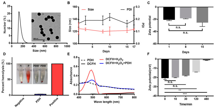
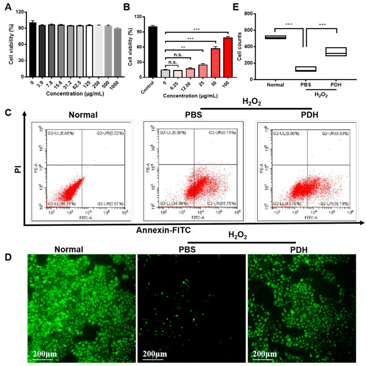
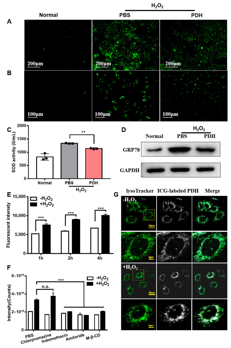
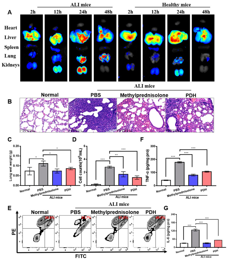
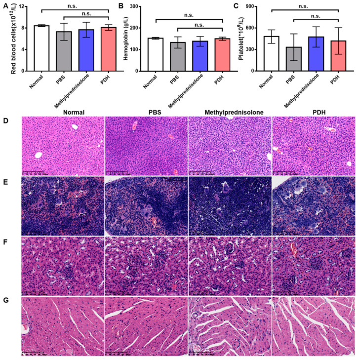
Nanoenzyme-mediated catalytic activity is emerging as a novel strategy for reactive oxygen species (ROS) scavenging in acute lung injury (ALI) treatment. However, one of the main hurdles for these metal-containing nanoenzymes is their potential toxicity and single therapeutic mechanism. Herein, we uncovered a melanin-like nanoparticles derived from the self-polymerization of 1,8-dihydroxynaphthalene (PDH nanoparticles), showing a significant anti-inflammation therapeutic effect on ALI mice. The prepared PDH nanoparticles rich in phenol groups could not only act as radical scavengers to alleviate oxidative stress but could also chelate calcium overload to suppress the endoplasmic reticulum stress response. As revealed by the therapeutic effect in vivo, PDH nanoparticles significantly prohibited neutrophil infiltration and the secretion of proinflammatory cytokines (TNF-α and IL-6), thus improving the inflammatory cascade in the ALI model. Above all, our work provides an effective anti-inflammatory nanoplatform by using the inherent capability of melanin-like nanoenzymes, proposing the potential application prospects of these melanin-like nanoparticles for acute inflammation-induced injury treatment.

Keywords: 1,8-DHN polymerized nanoparticles; acute lung injury; endoplasmic reticulum stress; melanin-like nanoenzyme; oxidative stress.


Conflict of interest statement

All authors declare no financial/commercial conflict of interest.

Figures

Figure 1
Figure 1
Characterization of PDH nanoparticles. (A) The size distribution and TEM images (inserted) of PDH nanoparticles. (B) Changes in nanoparticle size and polydispersity index after storage for 1, 3, 8, 10, 15, and 17 days. (C) Changes in zeta potential after storage for 1, 8, and 15 days. (D) The hemolysis rate and images (inserted) of red blood cells after incubation with different nanoparticles. N: negative control, P: positive control, PDH’: PDH sample mixed with saline, PDH: PDH sample mixed with red blood cells. (E) The absorbance of PDH, DCFH, DCFH + H2O2, and DCFH + H2O2 + PDH under different wavelengths. (F) Changes in zeta potential of PDH nanoparticles after incubation with calcium ion-containing solution (calcium chloride aqueous solution). (Results are presented as mean ± SD, n.s. is non-significant, ** p < 0.01, *** p < 0.001, n = 3).
Figure 2
Figure 2
In vitro therapeutic effect of PDH nanoparticles. (A) The cell viability of PDH nanoparticles in normal HUVECs detected by MTT assay. (B) The cell viability after treatment with different concentrations of PDH nanoparticles detected by MTT assay. (C) The annexin-FITC and PI staining of HUVECs after different treatments evaluated by flow cytometry. The first quadrant indicates living cells. The second quadrant indicates early apoptotic cells. The third quadrant represents late apoptotic cells. The fourth quadrant shows the necrotic cells. (D) Living cells (labeled with calcein AM) after different treatment with PDH. (E) Cell count in Figure 2D measured by image J. (n.s. is non-significant, ** p < 0.01, *** p < 0.001, n = 3).
Figure 3
Figure 3
In vitro therapeutic mechanism of PDH nanoparticles (50 μg/mL). (A) The ROS levels in cells after different treatments using the DCFH-DA probe. (B) The concentrations of free calcium ions in cells after different treatments detected with Fluo-4 AM probe. (C) The SOD activity in cells after different treatments was detected by total SOD assay kit with WST-8. Three circles in the figure mean three samples detected in the normal group. Three squares indicate three samples investigated in the PBS group. And three triangles represent three samples detected in the PDH group. (D) The expression levels of GRP78 in cells after different treatments were analyzed by Western blot. (E) The fluorescent intensities in cells treated with ICG-labeled PDH nanoparticles in the presence or absence of H2O2 for 1 h, 2 h, and 4 h were measured by flow cytometry. (F) The fluorescent intensity at 4 h in cells pretreated with different internalization inhibitors was measured by flow cytometry. (G) The co-localization of the lysosome tracker and ICG-labeled PDH nanoparticles at 4 h. (n.s. is non-significant, ** p < 0.01, *** p < 0.001, n = 3).
Figure 4
Figure 4
In vivo therapeutic effect of PDH nanoparticles on ALI-induced mice. (A) Biodistribution of PDH/ICG in vivo. The fluorescence images of harvested organs from mice treated with LPS or not at 2 h, 12 h, 24 h and 48 h. (B) Morphologic alterations in lungs after different treatments were identified by hematoxylin and eosin staining. Scale bar = 200 µm. (C) The lung wet weight. (D) The total cell counts in the bronchoalveolar lavage fluid detected by flow cytometry. (E) The neutrophil counts in the bronchoalveolar lavage fluid measured by flow cytometry. (F) The level of TNF-α in lung tissues detected by TNF-α ELISA kit. (G) The level of IL-6 in lung tissues detected by IL-6 ELISA kit. (* p < 0.05, ** p < 0.01, *** p < 0.001, n = 3).
Figure 5
Figure 5
The biosafety of PDH nanoparticles in ALI mice. The changes in red blood cells (A), hemoglobin (B), and platelets (C) detected by automatic blood analyzer (n.s. is non-significant, n = 6). Representative images of liver ((D), scale bar = 200 μm), spleen ((E), scale bar = 100 μm), kidney ((F), scale bar = 100 μm), and heart ((G), scale bar = 100 μm) identified with hematoxylin and eosin staining.

Similar articles

Cited by

References

    1. Gouda M.M., Bhandary Y.P. Acute Lung Injury: IL-17A-Mediated Inflammatory Pathway and Its Regulation by Curcumin. Inflammation. 2019;42:1160–1169. doi: 10.1007/s10753-019-01010-4. - DOI - PubMed
    1. Mowery N.T., Terzian W.T.H., Nelson A.C. Acute Lung Injury. Curr. Prob. Surg. 2020;57:100777. doi: 10.1016/j.cpsurg.2020.100777. - DOI - PubMed
    1. Butt Y., Kurdowska A., Allen T.C. Acute Lung Injury: A Clinical and Molecular Review. Arch. Pathol. Lab. Med. 2016;140:345–350. doi: 10.5858/arpa.2015-0519-RA. - DOI - PubMed
    1. Chacko B., Peter J.V., Tharyan P., John G., Jeyaseelan L. Pressure-controlled versus volume-controlled ventilation for acute respiratory failure due to acute lung injury (ALI) or acute respiratory distress syndrome (ARDS) Cochrane Database Syst. Rev. 2015;1:CD008807. doi: 10.1002/14651858.CD008807.pub2. - DOI - PMC - PubMed
    1. Kuldanek S.A., Kelher M., Silliman C.C. Risk factors, management and prevention of transfusion-related acute lung injury: A comprehensive update. Expert Rev. Hematol. 2019;12:773–785. doi: 10.1080/17474086.2019.1640599. - DOI - PMC - PubMed