. 2021 Nov 3;13(11):1851.
doi: 10.3390/pharmaceutics13111851.

Aerosol Delivery of Surfactant Liposomes for Management of Pulmonary Fibrosis: An Approach Supporting Pulmonary Mechanics

Affiliations

Aerosol Delivery of Surfactant Liposomes for Management of Pulmonary Fibrosis: An Approach Supporting Pulmonary Mechanics

Sabna Kotta et al. Pharmaceutics. .

Abstract

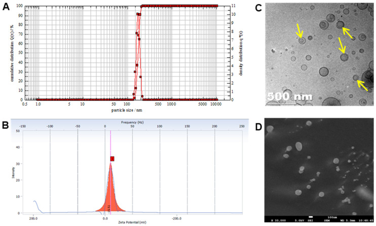
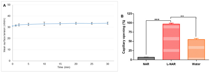
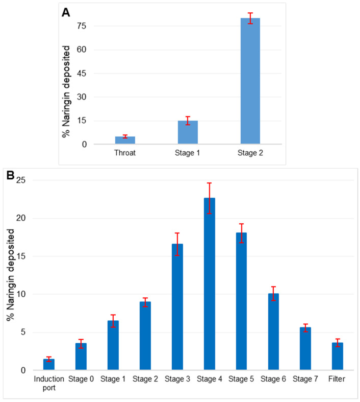
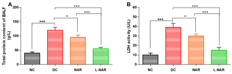
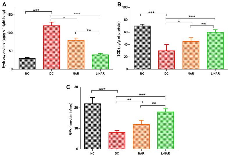
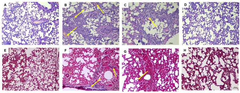
Excessive architectural re-modeling of tissues in pulmonary fibrosis due to proliferation of myofibroblasts and deposition of extracellular matrix adversely affects the elasticity of the alveoli and lung function. Progressively destructive chronic inflammatory disease, therefore, necessitates safe and effective non-invasive airway delivery that can reach deep alveoli, restore the surfactant function and reduce oxidative stress. We designed an endogenous surfactant-based liposomal delivery system of naringin to be delivered as an aerosol that supports pulmonary mechanics for the management of pulmonary fibrosis. Phosphatidylcholine-based liposomes showed 91.5 ± 2.4% encapsulation of naringin, with a mean size of 171.4 ± 5.8 nm and zeta potential of -15.5 ± 1.3 mV. Liposomes with the unilamellar structure were found to be spherical and homogeneous in shape using electron microscope imaging. The formulation showed surface tension of 32.6 ± 0.96 mN/m and was able to maintain airway patency of 97 ± 2.5% for a 120 s test period ensuring the effective opening of lung capillaries and deep lung delivery. In vitro lung deposition utilizing Twin Stage Impinger showed 79 ± 1.5% deposition in lower airways, and Anderson Cascade Impactor deposition revealed a mass median aerodynamic diameter of 2.35 ± 1.02 μm for the aerosolized formulation. In vivo efficacy of the developed formulation was analyzed in bleomycin-induced lung fibrosis model in rats after administration by the inhalation route. Lactate dehydrogenase activity, total protein content, and inflammatory cell infiltration in broncho-alveolar lavage fluid were substantially reduced by liposomal naringin. Oxidative stress was minimized as observed from levels of antioxidant enzymes. Masson's Trichrome staining of lung tissue revealed significant amelioration of histological changes and lesser deposition of collagen. Overall results indicated the therapeutic potential of the developed non-invasive aerosol formulation for the effective management of pulmonary fibrosis.

Keywords: aerosol; bleomycin; liposomes; naringin; pulmonary fibrosis.


Conflict of interest statement

The authors declare no conflict of interest.

Figures

Figure 1
Figure 1
Characterization of liposomal naringin. (A) Particle size analysis, (B) Zeta potential, (C) TEM image with arrows indicating the lamellae of vesicles, and (D) SEM image.
Figure 2
Figure 2
(A) Surface tension-time isotherms indicating adsorption of liposomal naringin. Each value represents the mean of 3 determinations. (B) Capillary patency of pristine naringin (NAR), liposomal naringin (L-NAR), and water measured using capillary surfactometer. The data is presented as the average of three determinations. Error bars represent standard deviation. **, and *** show significant difference by Newman–Keuls analysis following ANOVA at 95 percent confidence level at p < 0.01 and p < 0.001, respectively.
Figure 3
Figure 3
(A) In vitro lung deposition using Twin Stage Impinger (TSI) depicting deposition in different parts. (B) Anderson Cascade Impactor (ACI) deposition pattern of liposomal naringin on various stages of impactor at a flow rate of 15 L/min. Data is represented as a mean of 3 determinations. Error bars indicate standard deviation.
Figure 4
Figure 4
Effect on inflammatory cell counts in the BALF of rats, normal control (NC), disease control (DC), inhalation therapy of pristine naringin (NAR), and liposomal naringin (L-NAR) on with pulmonary fibrosis induced using intratracheal bleomycin. Total cells (A), Neutrophils (B) and Lymphocytes (C). The results were given as the mean standard error of the mean (n = 12). **, and *** show significant difference by Newman–Keuls analysis following ANOVA at 95 percent confidence level at p < 0.01 and p < 0.001 respectively.
Figure 5
Figure 5
Total protein (A) and Lactate dehydrogenase (LDH) activity (B) in BALF, from various groups, namely normal control (NC), disease control (DC), inhalation therapy of pristine naringin (NAR), and liposomal naringin (L-NAR) (n = 12). *, and *** indicate significant difference by Newman–Keuls analysis after ANOVA at 95 percent confidence level at p < 0.05, and p < 0.001 respectively.
Figure 6
Figure 6
Hydroxyproline (A), Superoxide dismutase (SOD, (B)), and Glutathione peroxidase (GPx, (C)) activity from various groups only normal control (NC), disease control (DC), inhalation therapy of pristine naringin (NAR), and liposomal naringin (L-NAR) (n = 12). *, ** and *** indicate significant difference by Newman–Keuls analysis after ANOVA at 95 percent confidence level at p < 0.05, p < 0.01 and p < 0.001 respectively.
Figure 7
Figure 7
Effect of daily inhaling therapy of liposomal naringin evaluated utilizing lung histological changes using H and E staining (100×). (A) Normal control, the lung architecture is intact, with no indications of injury, (B) Disease control, showing moderate fibrotic changes, (C) Pristine naringin therapy revealing mild to moderate fibrotic changes, and (D) Liposomal naringin treatment with the restoration of normal lung architecture. Masson’s Trichrome staining was used to examine lung histological changes (100×). (E) The lack of blue staining in the normal control indicates the absence of collagen deposition. (F) Disease control demonstrates the blue color of collagen deposition. (G) Pristine naringin treatment revealed mild bluish coloration of collagen, and (H) Liposomal naringin treatment showed significantly lesser deposition of collagen.

Similar articles

Cited by

References

    1. Ivanova V., Garbuzenko O.B., Reuhl K.R., Reimer D.C., Pozharov V.P., Minko T. Inhalation treatment of pulmonary fibrosis by liposomal prostaglandin E2. Eur. J. Pharm. Biopharm. 2013;84:335–344. doi: 10.1016/j.ejpb.2012.11.023. - DOI - PMC - PubMed
    1. Wallace W.A.H., Fitch P.M., Simpson A.J., Howie S.E.M. Inflammation-associated remodelling and fibrosis in the lung–A process and an end point. Int. J. Exp. Pathol. 2007;88:103–110. doi: 10.1111/j.1365-2613.2006.00515.x. - DOI - PMC - PubMed
    1. Chennakesavulu S., Mishra A., Sudheer A., Sowmya C., Reddy C.S., Bhargav E. Pulmonary delivery of liposomal dry powder inhaler formulation for effective treatment of idiopathic pulmonary fibrosis. Asian J. Pharm. Sci. 2018;13:91–100. doi: 10.1016/j.ajps.2017.08.005. - DOI - PMC - PubMed
    1. Kotta S., Aldawsari H.M., Badr-Eldin S.M., Binmahfouz L.S., Bakhaidar R.B., Sreeharsha N., Nair A.B., Ramnarayanan C. Lung Targeted Lipopolymeric Microspheres of Dexamethasone for the Treatment of ARDS. Pharmaceutics. 2021;13:1347. doi: 10.3390/pharmaceutics13091347. - DOI - PMC - PubMed
    1. Joshi N., Shirsath N., Singh A., Joshi K.S., Banerjee R. Endogenous lung surfactant inspired pH responsive nanovesicle aerosols: Pulmonary compatible and site-specific drug delivery in lung metastases. Sci. Rep. 2014;4:7085. doi: 10.1038/srep07085. - DOI - PMC - PubMed

LinkOut - more resources