. 2021 Nov 10;13(11):1904.
doi: 10.3390/pharmaceutics13111904.

Selective CNS Targeting and Distribution with a Refined Region-Specific Intranasal Delivery Technique via the Olfactory Mucosa

Affiliations

Selective CNS Targeting and Distribution with a Refined Region-Specific Intranasal Delivery Technique via the Olfactory Mucosa

Frank Maigler et al. Pharmaceutics. .

Abstract

Intranasal drug delivery is a promising approach for the delivery of drugs to the CNS, but too heterogenous, unprecise delivery methods without standardization decrease the quality of many studies in rodents. Thus, the lack of a precise and region-specific application technique for mice is a major drawback. In this study, a previously developed catheter-based refined technique was validated against the conventional pipette-based method and used to specifically reach the olfactory or the respiratory nasal regions. This study successfully demonstrated region-specific administration at the olfactory mucosa resulting in over 20% of the administered fluorescein dose in the olfactory bulbs, and no peripheral bioactivity of insulin detemir and Fc-dependent uptake of two murine IgG1 (11C7 and P3X) along the olfactory pathway to cortex and hippocampus. An scFv of 11C7 showed hardly any uptake to the CNS. Elimination was dependent on the presence of the IgG's antigen. In summary, it was successfully demonstrated that region-specific intranasal administration via the olfactory region resulted in improved brain targeting and reduced peripheral targeting in mice. The data are discussed with regard to their clinical potential.

Keywords: CNS drug delivery; CNS targeting; biopharmaceuticals; nose to brain drug delivery; refined drug delivery; therapeutic antibodies.


Conflict of interest statement

The authors declare that the research was conducted in the absence of any commercial or financial relationships that could be construed as a potential conflict of interest.

Figures
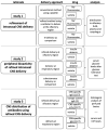
Figure 1
Figure 1
Study design of three studies with the aim of validating the previously refined intranasal region-specific administration technique [25] in vivo at the olfactory mucosa (study 1), determining the extent of molecules that are delivered to the periphery when administered at the olfactory mucosa (study 2) and analyzing the distribution of IgGs in the CNS after intranasal administration at the olfactory region (study 3).
Figure 2
Figure 2
The refined technique results in up to 21% of the dose in the olfactory bulbs, when administered at the olfactory region (study 1). (A) Sagittal view on a murine head with highlighted areas for catheter-based administration at either respiratory mucosa (RM) or olfactory mucosa (OM) with adjacent olfactory bulb (OB) as part of the brain. As previously described, a region-specific technique was developed with the aid of 3D-printed models of the murine nasal cavity reconstructed from CT scans of a mouse head [25]. (B) demonstrates the conventional technique using a pipette with usually a rather high volume, from 20 to 30 µL. With this method, no selective delivery to the respiratory or olfactory mucosa is possible. Therefore, the intranasal delivery (C) has been refined by using a catheter with an adjusted smaller administered volume, which can deliver the drug region-specific either at the olfactory or respiratory area. The aim of study 1 was to validate the region-specificity of the refined catheter-based technique with fluorescein in vivo. (D) 10 min after administration, the levels reached with the refined technique were significantly higher than achieved with the conventional method or IV delivery (* p < 0.04, Student’s t-test). At 30 min after administration at the olfactory region, 21.8 ± 5.34% of the dose was recovered at the olfactory bulbs while administration with a pipette at the nostrils resulted in only 1.5 ± 0.05% (n = 2; p = 0.0626) and intravenous (IV) delivery resulted in 0.7 ± 0.07% (n = 2; p = 0.0587). (E) Elevated levels of fluorescein were detected in serum after IV administration and after intranasal delivery with the conventional pipette-based method. (F) Interestingly, higher levels of traces of the intranasally administered doses were swallowed and ingested with the conventional method than with the refined method (conventional: 1.15 ± 0.804% vs. refined: 0.03 ± 0.078% vs. IV: 0.078 ± 0.078%). Due to the limited number of animals per group this study shows tendencies, but fails to demonstrate statistical significance (mean ± SEM; n = 2). (AC) are adapted with permission from ref. [25]. 2021, Nicole Lange et al.
Figure 3
Figure 3
No peripheral bioactivity after region-specific delivery of insulin detemir to the olfactory region (study 2). Silicone based 3D-model made by vacuum cast method (A), for the visualization of the targeted regions [25]. Adapted with permission from ref. [25]. 2021, Nicole Lange et al. Glucose tolerance test to examine the peripheral effects after region-specific administration of insulin detemir versus vehicle (B), monitored via blood glucose levels. Black arrows highlight catheter-based refined administration to the respiratory or olfactory nasal regions or subcutaneous injections of insulin detemir or vehicle. Mice were challenged with a high dose of intraperitoneal glucose to determine the peripheral activity of insulin detemir. As expected, subcutaneously delivered insulin demonstrated a high peripheral bioactivity by lowering the blood glucose levels. Interestingly, insulin delivered via the respiratory regions covered predominantly with respiratory mucosa was also distributed from the nose to the periphery and displayed 40.2% of the subcutaneously delivered activity. Insulin targeted to the olfactory regions did not show any statistically significant bioactivity in the periphery. Error bars represent mean ± SEM, n = 3. Data were analyzed with two-way ANOVA with multiple comparisons. ** p < 0.009. A is reproduced from [25] with kind permission from the rights owner.
Figure 4
Figure 4
Distribution of the CNS-specific IgG1 antibody 11C7 in the nasal mucosa after region-specific intranasal delivery (study 3). A total dose of 60 µg 11C7 per mouse was administered at the olfactory (BD,GI,LN) and respiratory (E,J,O) regions. The vehicle control was administered at the olfactory region and displays background staining from endogenous murine IgGs in the nasal mucus (A), and the choroid plexus (F), which was not removed after transcardial perfusion. The region-specificity became obvious in an investigation of the upper nasal cavity with its ethmoid turbinates: while after administration at the olfactory region the antibody was detectable at the olfactory mucosa (B), and showed a time-dependent clearance (C,D), no elevated levels of IgG were observed at the ethmoid turbinates after administration at the respiratory region. Further, none of the animals from the respiratory delivery group displayed any signs of a CNS delivery of 11C7 (J,O). A rapid distribution to the subventricular zones (see arrowheads) of the ventricles was found (G), that lasted up to 20 h (H), but was undetectable after 48 h (I). In addition, 11C7 was observed diffusely (see asterisks) in the hippocampus shortly after administration (L). The diffuse pattern disappeared within 20 h (M) and after 48 h distinct cells (see arrowheads) and neuronal projections were observed (N), highly similar to what was reported from Nogo-A expression studies [43]. Scale bar, 100 µm.
Figure 5
Figure 5
Transport mechanisms of both the full IgGs 11C7 and the isotype (IC) control, and the 11C7 scFv (study 3). Images from confocal microscopy show the rapid uptake of 11C7 and IC into the olfactory mucosa (OM) and also traces in the olfactory bulb (OB) after only 15 min (A,C). Higher magnification shows evidence for a predominantly intracellular pathway as previously reported from ex vivo olfactory mucosa (see arrowheads) (B,D) [38,39]. In contrast, the 11C7 scFv devoid of an Fc domain appears to be taken up to a lower extent (E) and displays more evidence for a transcellular transport (F). However, it should be noted that IgGs and scFv were visualized with different detections systems (anti-IgG vs. anti-His-Tag) and, hence, the fluorescence intensity should be evaluated with caution. Epiflourescence microscopy demonstrates, 45 min after administration, the full transport scheme with intracellular uptake at the apical mucosa, distribution to the lamina propria and transport along neuronal bundles from the lamina propria to the olfactory bulb (G). For a better visualization, anti-IgG immunoreactivity is displayed here in red, and neurofilament (NF200)-immunoreactivity in green. Transport kinetics along neuronal bundles after a single dose (HK): while only background is observed in the vehicle control animals (H), decreasing levels of 11C7 are observed within 48 h (IK). Arrowheads point to distinct stained structures while asterisk show diffuse staining pattern. Scale bar, 100 µm.
Figure 6
Figure 6
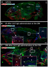
Time-dependent CNS distribution analyzed in sagittal section from mouse heads after region-specific delivery at the olfactory mucosa (OM, study 3). Whole slide images of a vehicle treated animal demonstrate endogenous IgGs in the nasal mucus, but also in the muscles underneath the nasal cavity and at the neck (A). This immunoreactivity was observed in all animals investigated. No background staining was observed in the CNS except for choroid plexus in all vehicle animals. The 11C7 could clearly be detected in the olfactory bulbs (arrowheads) and in the dentate gyrus of the hippocampus (B). At 20 h after administration the distribution pattern shifted into the caudal direction with lower intensities in the olfactory bulbs, but higher and more distinct staining in the cortex and the hippocampus where the antigen of 11C7 was expressed. Arrowheads point to distinct stained structures while asterisk show diffuse staining pattern. (C). OM, olfactory mucosa; RM, respiratory mucosa; OB, olfactory bulb; AON, anterior olfactory nucleus; DG, dentate gyrus. Scale bars, 1000 µm.
Figure 7
Figure 7
Antibody distribution is dependent on the presence of the antigen in the CNS and the presence of an Fc domain (study 3). The distribution and clearance in the murine OM and CNS are shown 45 min and 20 h (E,J,O,T) after directed intranasal administration at the olfactory region (OM). All images were captured with a confocal microscope. Arrowheads point to distinct stained structures while asterisk show diffuse staining pattern. (AE) Background signal due to endogenous IgGs, that have not been cleared by transcardial perfusion, and autofluorescence in the tissue of an animal, which received a vehicle control (PBS). (FJ) distribution of anti-Nogo-A monoclonal full antibody 11C7. The 11C7 was detected in the OM (F), the OB (G), the AON/OT (anterior olfactory nucleus/olfactory tubercle) (H), the choroid plexus in the ventricles (I), and after 20 h in the hippocampus (J). The strongest signal was detected in the olfactory epithelium followed by the olfactory bulb and the choroid plexus. In the hippocampus, the signals were amplified for a better visualization. (KO) Distribution of the isotype control (IC) antibody, which does not recognize any structure of the murine CNS as antigen. The distribution profile within 45 min was similar to 11C7 however the immunoreactivity in the AON/OT was lower in all animals investigated. The similar distribution pattern in OM, OB, AON and the subventricular zones imply the relevance of the Fc receptor system. The absence of IC in hippocampus (O) is a strong indication that the presence of the antigen is critical to avoid rapid elimination. (PT) A scFv format of 11C7 was distributed to the OB to a lesser extent than the IgGs. In addition, hardly any signals higher than the vehicle control could be found in the AON/OT, choroid plexus, subventricular zones, nor in the Nogo-A expressing cells in the hippocampus. Nevertheless, since the 11C7 scFv was detected via its penta His-Tag, the intensities of (AO) should not be directly compared with (PT). Representative images are shown. OM, olfactory mucosa; OB, olfactory bulb; AON, anterior olfactory nucleus; OT, olfactory tubercle. Scale bar: 100 µm.

Similar articles

Cited by

References

    1. Wang Z., Xiong G., Tsang W.C., Schätzlein A.G., Uchegbu I.F. Nose-to-brain delivery. J. Pharmacol. Exp. Ther. 2019;370:593–601. doi: 10.1124/jpet.119.258152. - DOI - PubMed
    1. Zhang Y., Chan H.F., Leong K.W. Advanced materials and processing for drug delivery: The past and the future. Adv. Drug Deliv. Rev. 2013;65:104–120. doi: 10.1016/j.addr.2012.10.003. - DOI - PMC - PubMed
    1. Neuwelt E., Abbott N.J., Abrey L., Banks W.A., Blakley B., Davis T., Engelhardt B., Grammas P., Nedergaard M., Nutt J. Strategies to advance translational research into brain barriers. Lancet Neurol. 2008;7:84–96. doi: 10.1016/S1474-4422(07)70326-5. - DOI - PubMed
    1. Dhuria S.V., Hanson L.R., Frey W.H. Intranasal delivery to the central nervous system: Mechanisms and experimental considerations. J. Pharm. Sci. 2010;99:1654–1673. doi: 10.1002/jps.21924. - DOI - PubMed
    1. Illum L. Transport of drugs from the nasal cavity to the central nervous system. Eur. J. Pharm. Sci. 2000;11:1–18. doi: 10.1016/S0928-0987(00)00087-7. - DOI - PubMed

LinkOut - more resources