. 2021 Nov 15;13(11):1929.
doi: 10.3390/pharmaceutics13111929.

Complementary Nucleobase Interactions Drive Co-Assembly of Drugs and Nanocarriers for Selective Cancer Chemotherapy

Affiliations

Complementary Nucleobase Interactions Drive Co-Assembly of Drugs and Nanocarriers for Selective Cancer Chemotherapy

Fasih Bintang Ilhami et al. Pharmaceutics. .

Abstract

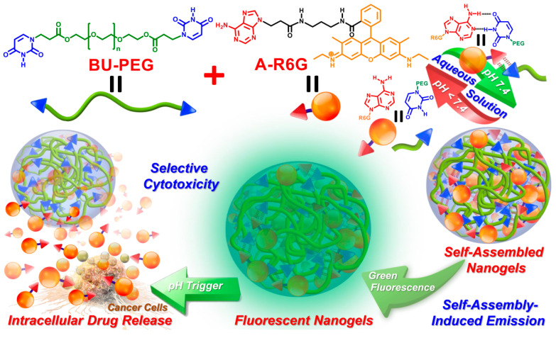
A new concept in cooperative adenine-uracil (A-U) hydrogen bonding interactions between anticancer drugs and nanocarrier complexes was successfully demonstrated by invoking the co-assembly of water soluble, uracil end-capped polyethylene glycol polymer (BU-PEG) upon association with the hydrophobic drug adenine-modified rhodamine (A-R6G). This concept holds promise as a smart and versatile drug delivery system for the achievement of targeted, more efficient cancer chemotherapy. Due to A-U base pairing between BU-PEG and A-R6G, BU-PEG has high tendency to interact with A-R6G, which leads to the formation of self-assembled A-R6G/BU-PEG nanogels in aqueous solution. The resulting nanogels exhibit a number of unique physical properties, including extremely high A-R6G-loading capacity, well-controlled, pH-triggered A-R6G release behavior, and excellent structural stability in biological media. Importantly, a series of in vitro cellular experiments clearly demonstrated that A-R6G/BU-PEG nanogels improved the selective uptake of A-R6G by cancer cells via endocytosis and promoted the intracellular release of A-R6G to subsequently induce apoptotic cell death, while control rhodamine/BU-PEG nanogels did not exert selective toxicity in cancer or normal cell lines. Overall, these results indicate that cooperative A-U base pairing within nanogels is a critical factor that improves selective drug uptake and effectively promotes apoptotic programmed cell death in cancer cells.

Keywords: adenine–uracil base pair; complementary hydrogen bonded drug carrier system; controlled drug delivery; selective cytotoxicity; supramolecular nanogels.


Conflict of interest statement

The authors declare no conflict of interest.

Figures

Scheme 1
Scheme 1
Schematic illustration of the co-assembly process, fluorescent properties, and cancer cell-selective cytotoxic behavior of hydrogen-bonded A-R6G/BU-PEG complexes. The upper-right green and red arrows represent the association and dissociation of the complementary A–U base pairs under various pH conditions.
Figure 1
Figure 1
(a) Determination of the CMC values of PEG and BU-PEG in water in the presence of pyrene. (b) Hydrodynamic particle size of the BU-PEG polymer in water as determined by DLS at 25 °C. Surface morphologies of spin-coated BU-PEG polymers obtained by (c) AFM and (d) SEM at 25 °C.
Figure 2
Figure 2
(a) AMF images of A-R6G-loaded BU-PEG nanogels measured at 25 °C. (b) PL spectra of A-R6G-loaded and R6G-loaded BU-PEG nanogels in water at 25 °C. Inset: photographs of A-R6G-loaded and R6G-loaded BU-PEG nanogels in water exposed to (1) natural lighting and (2) broadband UV lighting conditions. (c) Time-dependent DLS analysis of the structural stability of A-R6G-loaded and R6G-loaded BU-PEG nanogels in media containing 10% FBS at pH 7.4 for 24 h. (d) In vitro hemolytic assay of different concentrations of A-R6G-loaded and R6G-loaded BU-PEG nanogels on SRBCs. Inset: photographs showing SRBCs incubated with varying concentrations (1–100 μg/mL) of A-R6G-loaded or R6G-loaded BU-PEG nanogels at 37 °C in 5% CO2 for 4 h.
Figure 3
Figure 3
In vitro drug release profiles of (a) A-R6G-loaded and (b) R6G-loaded BU-PEG nanogels. In vitro cytotoxicity of A-R6G-loaded and R6G-loaded BU-PEG nanogels against (c) NIH/3T3 cells and (d) HeLa cells after 24 h incubation.
Figure 4
Figure 4
CLSM images of (a) NIH/3T3 cells and (b) HeLa cells cultured with A-R6G-loaded BU-PEG nanogels at normal physiological conditions (pH 7.4 and 37 °C) for 3, 12, or 24 h. The scale bars in all CLSM images are 20 µm.
Figure 5
Figure 5
Flow cytometry histograms of (a) NIH/3T3 cells and (b) HeLa cells cultured with A-R6G-loaded or R6G-loaded BU-PEG nanogels at normal physiological conditions (pH 7.4 and 37 °C) for 1, 3, 6, 12, or 24 h.
Figure 6
Figure 6
Flow cytometric dot plot quadrant charts of NIH/3T3 and HeLa cells cultured with A-R6G-loaded and R6G-loaded BU-PEG nanogels at normal physiological conditions (pH 7.4 and 37 °C) for 1, 3, 6, or 12 h before staining with BV421 Annexin V and GDR780. Figures (a)–(p) represent the results of flow cytometry for different time points during the co-culture period. The graph quadrants from the lower left to the upper left (turning anti-clockwise) represent viable cells (BV421 Annexin V-, GDR780), early apoptotic cells (BV421 Annexin V+, GDR780), late apoptotic cells (BV421 Annexin V+, GDR780+), and necrotic cells (BV421 Annexin V, GDR780+). The numbers inside each quadrant refer to the proportions of cells.

References

    1. Blanchard A.T., Salaita K. Emerging uses of DNA mechanical devices. Science. 2019;365:1080–1081. doi: 10.1126/science.aax3343. - DOI - PMC - PubMed
    1. Ouchi M., Badi N., Lutz J.-F., Sawamoto M. Single-chain technology using discrete synthetic macromolecules. Nat. Chem. 2011;3:917–924. doi: 10.1038/nchem.1175. - DOI - PubMed
    1. Badi N., Lutz J.-F. Sequence control in polymer synthesis. Chem. Soc. Rev. 2009;38:3383–3390. doi: 10.1039/b806413j. - DOI - PubMed
    1. Merrifield R.B. Solid phase peptide synthesis. I. The synthesis of a tetrapeptide. J. Am. Chem. Soc. 1963;85:2149–2154. doi: 10.1021/ja00897a025. - DOI
    1. Müller A., Talbot F., Leutwyler S. Hydrogen bond vibrations of 2-aminopyridine·2-pyridone, a Watson−Crick analogue of adenine·uracil. J. Am. Chem. Soc. 2002;124:14486–14494. doi: 10.1021/ja0209969. - DOI - PubMed

LinkOut - more resources