. 2021 Oct 27;10(11):2313.
doi: 10.3390/plants10112313.

Morphology and Anatomy of Branch-Branch Junctions in Opuntia ficus-indica and Cylindropuntia bigelovii: A Comparative Study Supported by Mechanical Tissue Quantification

Affiliations

Morphology and Anatomy of Branch-Branch Junctions in Opuntia ficus-indica and Cylindropuntia bigelovii: A Comparative Study Supported by Mechanical Tissue Quantification

Max D Mylo et al. Plants (Basel). .

Abstract

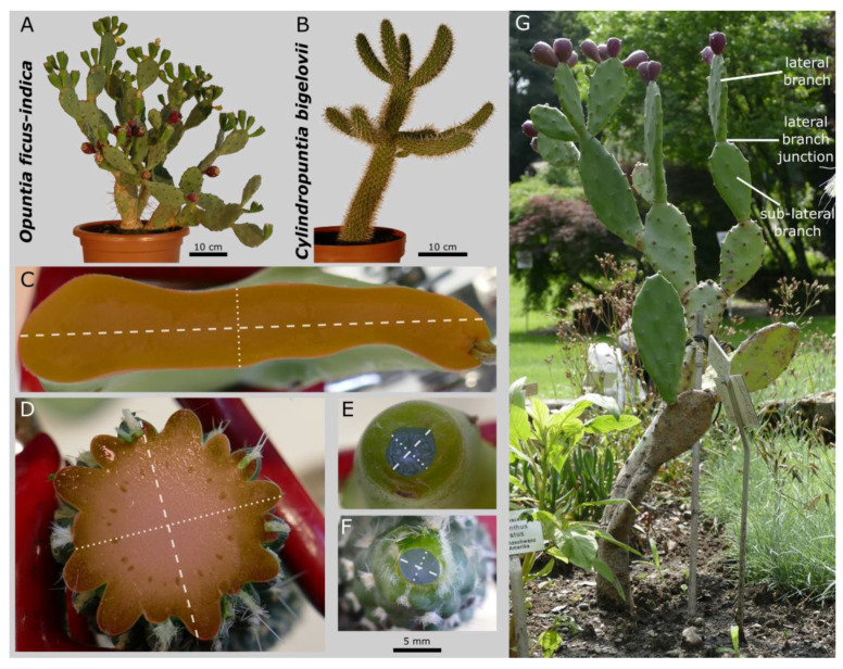
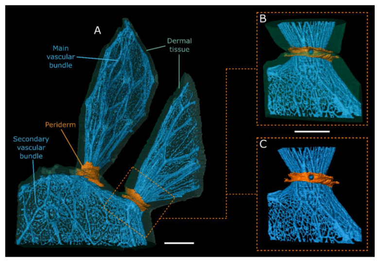
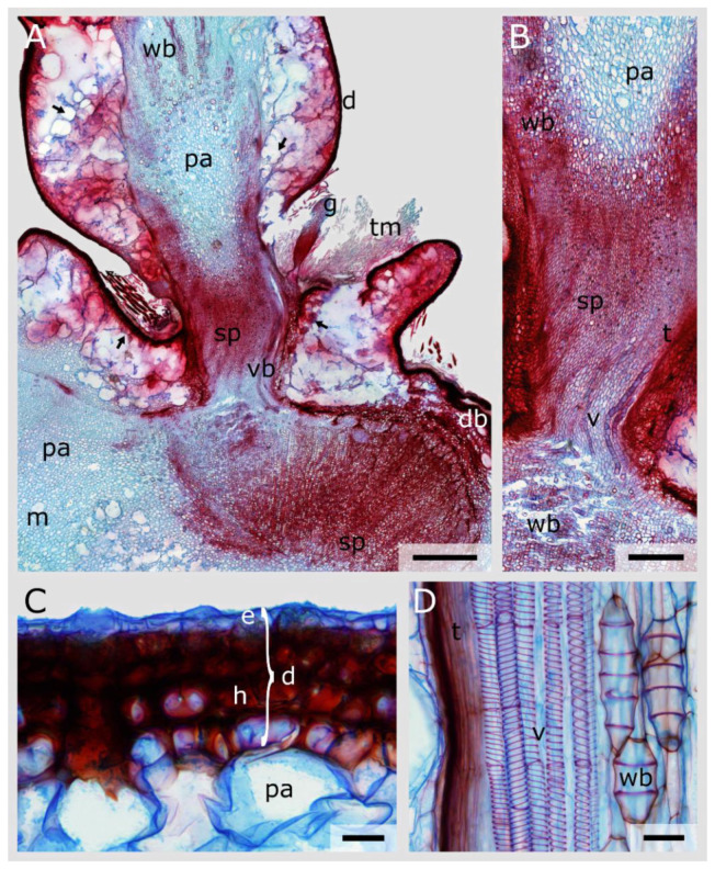
The Opuntioideae include iconic cacti whose lateral branch-branch junctions are intriguing objects from a mechanical viewpoint. We have compared Opuntia ficus-indica, which has stable branch connections, with Cylindropuntia bigelovii, whose side branches abscise under slight mechanical stress. To determine the underlying structures and mechanical characteristics of these stable versus shedding cacti junctions, we conducted magnetic resonance imaging, morphometric and anatomical analyses of the branches and tensile tests of individual tissues. The comparison revealed differences in geometry, shape and material properties as follows: (i) a more pronounced tapering of the cross-sectional area towards the junctions supports the abscission of young branches of C. bigelovii. (ii) Older branches of O. ficus-indica form, initially around the branch-branch junctions, collar-shaped periderm tissue. This secondary coverage mechanically stiffens the dermal tissue, giving a threefold increase in strength and a tenfold increase in the elastic modulus compared with the epidermis. (iii) An approximately 200-fold higher elastic modulus of the vascular bundles of O. ficus-indica is a prerequisite for the stable junction of its young branches. Our results provide, for both biological and engineered materials systems, important insights into the geometric characteristics and mechanical properties of branching joints that are either stable or easily detachable.

Keywords: Opuntioideae; abscission; cacti; magnetic resonance imaging; periderm formation; tissue tensile testing.


Conflict of interest statement

The authors declare no conflict of interest.

Figures

Figure 1
Figure 1
Experimental plants of Opuntia ficus-indica (A) and Cylindropuntia bigelovii (B) and their branch and junction cross-sections (O. ficus-indica: (C,E) and C. bigelovii: (D,F)). Exemplary representation of the maximum cross-sectional areas of the branches ((C,D); coloured red; areoles left out) and the areas of the junction zone ((E,F); coloured blue). The major axis (dashed white line; defined as the longest axis through the centroid) and the minor axis (dotted white line; orthogonal to the major axis through the centroid) of the respective cross-section are marked. The scale bar applies to subfigures (CF). An Opuntia plant from the Botanic Garden Freiburg (not used as the experimental plant) used to indicate the lateral and sub-lateral branches and the lateral branch junction (G).
Figure 2
Figure 2
MRI overview scans with exemplary segmentation of the respective slices. Tissue segmentation based on the grey-value signal of the MRI overview scans. Pseudo colour visualisation of the tissues of Opuntia ficus-indica (AC) and Cylindropuntia bigelovii (DF) including two sectional planes each ((B,E): tangential; (C,F): transverse) for exemplary visualisation of the raw MRI data including segmented areas. d: dermal tissue (green); m: mucilage cell or channel (not segmented); o: oxalate crystal (not segmented); p: peridermal tissue (orange; only in (A,B)); v: vascular structures (blue). White arrows exemplarily show the locations of the areoles. All scale bars equal 1 cm.
Figure 3
Figure 3
MRI scans of Opuntia ficus-indica. 3D representation of the dermal tissue ((A,B); transparent green), the vascular system ((AC); blue) and the peridermal tissue ((AC); orange) of a junction site and parts of the connected lateral and sub-lateral branches, based on the segmentation of MRI scans. (A) Visualisation (lateral view) of an overview scan (sample 1). Please note that the right and lower branch had to be trimmed because of space limitations of the MRI scanner. Visualisation (lateral view) of the surface coil scan (sample 2) with (B) and without (C) representation of dermal tissue. All scale bars equal 1 cm.
Figure 4
Figure 4
MRI scans of Cylindropuntia bigelovii. 3D representation of the dermal tissue (A); transparent green) and the vascular system ((AD); coloured blue) of the junctions and parts of the connected lateral and sub-lateral branches, based on the segmentation of MRI scans. (A,B) Visualisation (lateral view) of an overview scan (sample 1). (C,D) Visualisation of a surface coil scan (sample 2) with a distinction between vascular structures that supply the lateral branch (dark blue) and those that run in a dormant bud (light blue). All scale bars equal 1 cm.
Figure 5
Figure 5
Stained longitudinal microscopic thin sections of a lateral junction of Opuntia ficus-indica. (A) Overview image showing the vascular bundles (vb), the transition of the parenchyma (pa) to small-volume parenchyma cells (sp) and the peridermal coverage (pe) of the dermal tissue (d). The arrows indicate locations where the periderm detached during preparation of the sections. Trichomes (tm) are visible in the notch of the junction. (B) Detailed image of the junction area with small-volume parenchyma (sp) and vascular bundles consisting of vessel elements (v) and tracheids (t). (C) Detailed image of tracheids (t), vessels with mostly annular secondary thickening of the cell wall (v) and the less frequent wide-band tracheids (wb) embedded in the large-lumened parenchymatous tissue (pa) and located apical to the junction. (D) Detailed image of the dermal tissue (d), consisting of a single-layered epidermis (e), which is covered by a cuticle, and a multi-layered hypodermis (h), which is partly covered by peridermal layers (pe), consisting of densely packed phellem cells (ph), a one-layered meristematic phellogen (pg) and thin-walled phelloderm cells (pd). Stomata (s) and crystalline inclusions (black, circular structures beneath the epidermal layers) are visible. Scale bars: (A) 2 mm, (BD) 1000 µm.
Figure 6
Figure 6
Stained longitudinal microscopic thin sections of a lateral junction of Cylindropuntia bigelovii. (A) Overview image showing the vascular bundles (vb) partly running through the junction or running into a dormant bud (db), the transition of the parenchyma cells (pa and sp), the dermal tissue (d) and the mucilage cells (m). The arrows indicate locations where the dermal tissue detached from the cortical parenchyma during preparation of the sections. Trichomes (tm) and a glochid (g) are visible in the wrinkle of the junction. (B) Detailed image of the junction and the transition between the pith parenchyma (pa) and the small-volume parenchyma (sp). Tracheids (t) and vessel elements (v) run through the junction, whereas wide-band tracheids (wb) are present laterally and basally. (C) Detailed image of the dermal tissue (d), consisting of a single layered epidermis (e) covered by a thin cuticle and of a multi-layered hypodermis (h). (D) Detailed image of vessel elements with spiral secondary thickening of the cell wall (v), tracheids (t) and wide-band tracheids (wb) embedded in parenchymatous tissue. Scale bars: (A) 2 mm, (B) 1 mm, (C,D) 50 µm.
Figure 7
Figure 7
Stained transverse microscopic serial sections of a lateral junction of Opuntia ficus-indica. (AJ) Sections sorted from apical (A) to basal (J) in the junction, as schematically shown in (K). The scale bar applies to subfigures (AJ). a: areolar protrusion; c: cortex, consisting of parenchyma cells; d: dermal tissue, consisting of an epidermis covered with a cuticle and a hypodermis; m: mucilage channel; oc: outgrowing cavity; ov: outgrowing vascular bundle; p: pith, consisting of parenchyma cells of different sizes and with different cell wall thicknesses; pe: periderm; vb: vascular bundle. (L,M) Detailed images of the vascular bundles within the junction (L) and apical to it (M). v: vessel element; t: tracheid; wb: wide-band tracheid. (N) Detailed image of an outgrowing cavity (oc) with trichomes (tm).

References

    1. Nobel P.S. Cacti: Biology and Uses. University of California Press; Berkeley, CA, USA: 2002.
    1. Benson L. The Cacti of the United States and Canada. Stanford University Press; Stanford, CA, USA: 1982.
    1. Griffith M.P., Porter J.M. Phylogeny of Opuntioideae (Cactaceae) Int. J. Plant Sci. 2009;170:107–116. doi: 10.1086/593048. - DOI
    1. Mauseth J.D. Structure-function relationships in highly modified shoots of Cactaceae. Ann. Bot. 2006;98:901–926. doi: 10.1093/aob/mcl133. - DOI - PMC - PubMed
    1. Conde L.F. Anatomical Comparisons of Five Species of Opuntia (Cactaceae) Ann. Mo. Bot. Gard. 1975;62:425. doi: 10.2307/2395206. - DOI

LinkOut - more resources