. 2021 Oct 21;13(11):2125.
doi: 10.3390/v13112125.

Genotypic Variants of Pandemic H1N1 Influenza A Viruses Isolated from Severe Acute Respiratory Infections in Ukraine during the 2015/16 Influenza Season

Affiliations

Genotypic Variants of Pandemic H1N1 Influenza A Viruses Isolated from Severe Acute Respiratory Infections in Ukraine during the 2015/16 Influenza Season

Oksana Zolotarova et al. Viruses. .

Abstract

Human type A influenza viruses A(H1N1)pdm09 have caused seasonal epidemics of influenza since the 2009-2010 pandemic. A(H1N1)pdm09 viruses had a leading role in the severe epidemic season of 2015/16 in the Northern Hemisphere and caused a high incidence of acute respiratory infection (ARI) in Ukraine. Serious complications of influenza-associated severe ARI (SARI) were observed in the very young and individuals at increased risk, and 391 fatal cases occurred in the 2015/16 epidemic season. We analyzed the genetic changes in the genomes of A(H1N1)pdm09 influenza viruses isolated from SARI cases in Ukraine during the 2015/16 season. The viral hemagglutinin (HA) fell in H1 group 6B.1 for all but four isolates, with known mutations affecting glycosylation, the Sa antigenic site (S162N in all 6B.1 isolates), or virulence (D222G/N in two isolates). Other mutations occurred in antigenic site Ca (A141P and S236P), and a subgroup of four strains were in group 6B.2, with potential alterations to antigenicity in A(H1N1)pdm09 viruses circulating in 2015/16 in Ukraine. A cluster of Ukrainian isolates exhibited novel D2E and N48S mutations in the RNA binding domain, and E125D in the effector domain, of immune evasion nonstructural protein 1 (NS1). The diverse spectrum of amino-acid substitutions in HA, NS1, and other viral proteins including nucleoprotein (NP) and the polymerase complex suggested the concurrent circulation of multiple lineages of A(H1N1)pdm09 influenza viruses in the human population in Ukraine, a country with low vaccination coverage, complicating public health measures against influenza.

Keywords: A(H1N1)pdm09; H1N1; acute respiratory infection; influenza; mutation; phylogenetics.


Conflict of interest statement

The authors declare no conflict of interest.

Figures

Figure 1
Figure 1
Weekly confirmed cases of influenza in the 2013/14, 2014/15, and 2015/16 seasons in Ukraine among clinical surveillance for ILI in four cities in Ukraine (Kyiv, Dnipro, Odessa, and Khmelnytsky). Influenza type and subtype were determined by RT-PCR and antigenic testing from nasal swabs/sputum.
Figure 2
Figure 2
Phylogenetic analysis of A(H1N1)pdm09 influenza virus hemagglutinin (HA) nucleotide sequences from the 2015/16 season in Ukraine. Phylogenetic trees were constructed by the NJ (neighbor-joining) method, and Kimura 2-parameter model, with 1000 bootstrap replications. Amino acid variations are labeled for 6B.1 and 6B.2 subgroups.
Figure 3
Figure 3
Model 3D structure of the A(H1N1)pdm09 HA protein with the substitutions that occurred in 2015/16 in strains isolated in Ukraine. Structure (PDB ID—3LZG) was modeled using Chimera software and labeled according to H1 6B.1 (blue) or 6B.2 (orange) genetic group.
Figure 4
Figure 4
3D structure of HA molecule with changes in antigenic sites of Ukrainian isolates (PDB ID—3LZG).
Figure 5
Figure 5
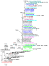
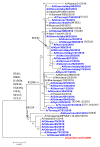
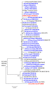
Phylogenetic comparison of A(H1N1)pdm09 influenza virus NA from the 2015/16 season. NA nucleotide sequences were used to build a tree by NJ method, Kimura 2-parameter model, with 1000 bootstrap replications. Amino acid variations are indicated; strains from Ukraine are colored; reference strains are black, and the vaccine strain is red.
Figure 6
Figure 6
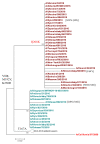
Phylogenetic analysis of A(H1N1)pdm09 influenza virus M1 from the 2015/16 season. M1 gene nucleotide sequences were analyzed by NJ method, Kimura 2-parameter model, with 1000 bootstrap replications. Ukrainian clades are colored except the vaccine strain which is red; amino acid variations are indicated as arising at specific branch nodes; M2 substitutions are also annotated (M2).
Figure 7
Figure 7
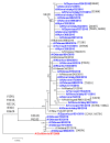
Phylogenetic comparison of A(H1N1)pdm09 influenza virus NP from the 2015/16 season. NP nucleotide sequences were analyzed by NJ method, Kimura 2-parameter model, with 1000 bootstrap replications. Ukrainian clades are colored blue; reference strains are black, except the vaccine strain (red/bold); amino acid variations are indicated, with M195T common in all isolates (red).
Figure 8
Figure 8
3D structure of NS1 molecule with changes in the Ukrainian isolates (PDB ID—3F5T). New mutations of NS1 of A(H1N1)pdm09 are shown in red. (Chimaera).
Figure 9
Figure 9
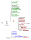
Phylogenetic comparison of A(H1N1)pdm09 influenza virus NS1 from the 2015/16 season. NS1 nucleotide sequences were analyzed by NJ method, Kimura 2-parameter model, with 1000 bootstrap replications. The Ukrainian 2014/15 subgroup (green) was distinct from 2015/16 isolates (brown) and a separate subgroup (blue); reference strains are black, except the vaccine strain (red/bold).
Figure 10
Figure 10
Phylogenetic comparison of A(H1N1)pdm09 influenza virus PA from the 2015/16 season. PA nucleotide sequences were analyzed by NJ method, Kimura 2-parameter model, with 1000 bootstrap replications. The Ukrainian 2015/16 isolates (blue), reference strains (black), and the vaccine strain (red/bold).
Figure 11
Figure 11
Phylogenetic comparison of A(H1N1)pdm09 influenza virus PB2 from the 2015/16 season. PB2 nucleotide sequences were analyzed by NJ method, Kimura 2-parameter model, with 1000 bootstrap replications. The Ukrainian 2015/16 isolates (blue), reference strains (black), and the vaccine strain (red/bold).
Figure 12
Figure 12
Phylogenetic comparison of A(H1N1)pdm09 influenza virus PB1 from the 2015/16 season. PB1 nucleotide sequences were analyzed by NJ method, Kimura 2-parameter model, with 1000 bootstrap replications. The Ukrainian 2015/16 isolates (blue), reference strains (black), and the vaccine strain (red/bold).

References

    1. Nelson M., Spiro D., Wentworth D., Beck E., Fan J., Ghedin E., Halpin R., Bera J., Hine E., Proudfoot K., et al. The early diversification of influenza A/H1N1pdm. PLoS Curr. Influenza. 2009;1:RRN1126. doi: 10.1371/currents.RRN1126. - DOI - PMC - PubMed
    1. Schweiger B., Zadow I., Heckler R. Antigenic drift and variability of influenza viruses. Med. Microbiol. Immunol. 2002;191:133–138. doi: 10.1007/s00430-002-0132-3. - DOI - PubMed
    1. Smith D., Lapedes A., de Jong J., Bestebroer T., Rimmelzwaan G., Osterhaus A., Fouchier R. Mapping the antigenic and genetic evolution of influenza virus. Science. 2004;305:371–376. doi: 10.1126/science.1097211. - DOI - PubMed
    1. Erbelding E., Post D., Stemmy E., Roberts P., Augustine A., Ferguson S., Paules C., Graham B., Fauci A. A Universal Influenza Vaccine: The Strategic Plan for the National Institute of Allergy and Infectious Diseases. J. Infect. Dis. 2018;218:347–354. doi: 10.1093/infdis/jiy103. - DOI - PMC - PubMed
    1. Belanov S., Bychkov D., Benner C., Ripatti S., Ojala T., Kankainen M., Lee H., Tang J., Kainov D. Genome-Wide Analysis of Evolutionary Markers of Human Influenza A(H1N1)pdm09 and A(H3N2) Viruses May Guide Selection of Vaccine Strain Candidates. Genome Biol. Evol. 2015;7:3472–3483. doi: 10.1093/gbe/evv240. - DOI - PMC - PubMed

Publication types

MeSH terms

Substances