Review
. 2021 Oct 29;13(11):2188.
doi: 10.3390/v13112188.

Interspecies Jumping of Bat Coronaviruses

Affiliations
Review

Interspecies Jumping of Bat Coronaviruses

Antonio C P Wong et al. Viruses. .

Abstract

In the last two decades, several coronavirus (CoV) interspecies jumping events have occurred between bats and other animals/humans, leading to major epidemics/pandemics and high fatalities. The SARS epidemic in 2002/2003 had a ~10% fatality. The discovery of SARS-related CoVs in horseshoe bats and civets and genomic studies have confirmed bat-to-civet-to-human transmission. The MERS epidemic that emerged in 2012 had a ~35% mortality, with dromedaries as the reservoir. Although CoVs with the same genome organization (e.g., Tylonycteris BatCoV HKU4 and Pipistrellus BatCoV HKU5) were also detected in bats, there is still a phylogenetic gap between these bat CoVs and MERS-CoV. In 2016, 10 years after the discovery of Rhinolophus BatCoV HKU2 in Chinese horseshoe bats, fatal swine disease outbreaks caused by this virus were reported in southern China. In late 2019, an outbreak of pneumonia emerged in Wuhan, China, and rapidly spread globally, leading to >4,000,000 fatalities so far. Although the genome of SARS-CoV-2 is highly similar to that of SARS-CoV, patient zero and the original source of the pandemic are still unknown. To protect humans from future public health threats, measures should be taken to monitor and reduce the chance of interspecies jumping events, either occurring naturally or through recombineering experiments.

Keywords: COVID-19; MERS; SADS; SARS; bat; coronavirus; epidemic; interspecies jumping; outbreak; pandemic.


Conflict of interest statement

The authors declare no conflict of interest.

Figures

Figure 1
Figure 1
Overview of interspecies transmission events of SARS epidemic in 2003 and genome organizations of SARSr-CoV from different hosts. Arrows indicate the direction of viral interspecies jumping and viral molecular evolution between different host species. Circular arrows indicate the circulation of virus within the population. ORF1a and ORF1b are represented by blue boxes, structural proteins by pink boxes, and accessory proteins by yellow boxes.
Figure 2
Figure 2
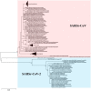
Maximum-likelihood phylogeny based on the nsp12 amino acid sequences of selected SARSr-CoVs. A Jones–Taylor–Thornton (JTT) model of amino acid substitution was used in the analysis with a discrete gamma (γ) distribution and the assumption that a certain fraction of sites is evolutionarily invariable (+I). 1000 trees were set for bootstrap values calculation. All bootstrap values and the scale bar indicating the number of amino acid substitutions per site are shown. Viral strains highlighted in red color represents the SARSr-CoV cluster; blue color represents the SARSr-CoV-2 cluster.
Figure 3
Figure 3
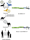
Overview of interspecies transmission events of MERS epidemic since 2012. Arrows indicate the direction of viral interspecies jumping between different host species. Circular arrows indicate the circulation of virus within the population. Dot arrows indicate the viral interspecies jumping event remains to be elucidated. ORF1a and ORF1b are represented by green boxes, structural proteins by blue and purple boxes, and accessory proteins by yellow boxes.
Figure 4
Figure 4
Maximum-likelihood phylogeny based on the nsp12 amino acid sequences of selected MERSr-CoVs and merbecoviruses. A Jones–Taylor–Thornton (JTT) model of amino acid substitution was used in the analysis with a discrete gamma (γ) distribution. 1000 trees were set for bootstrap values calculation. All bootstrap values and the scale bar indicating the number of amino acid substitutions per site are shown. Viral strains highlighted in red color represents the MERSr-CoV cluster, blue color represents the bat merbecoviruses cluster, yellow color represents the hedgehog merbecoviruses cluster.
Figure 5
Figure 5
Overview of interspecies transmission events of swine-related BatCoV HKU2 (SADS) outbreak since 2017. Arrows indicate the direction of viral interspecies jumping between different host species. Circular arrows indicate the circulation of virus within the population. Dot arrows indicate the viral interspecies jumping event remains to be elucidated.
Figure 6
Figure 6
Maximum-likelihood phylogeny based on the amino acid sequences of selected BatCoV HKU2 and swine-related BatCoV HKU2 (a) nsp12 and (b) S1 domains. (a) a Jones–Taylor–Thornton (JTT) model of amino acid substitution and (b) a Whelan–Goldman matrix (WAG) model with a discrete gamma (γ) distribution of amino acid substitution was used in the respective analyses. 1000 trees were set for bootstrap values calculation. All bootstrap values and the scale bars indicating the number of amino acid substitutions per site are shown. Viral strains highlighted in pink color represents Table 2 cluster; brown color represents the BatCoV HKU2 cluster.
Figure 7
Figure 7
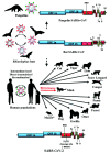
Overview of interspecies transmission events of COVID-19 pandemic since 2019. Arrows indicate the direction of viral interspecies jumping and viral molecular evolution between different host species. Circular arrows indicate the circulation of virus within the population. Dot arrows indicate the viral interspecies jumping event remains to be elucidated. Red arrows indicate the bi-directionality of interspecies jumping events. Asterisk labeled animal represents the laboratory interspecies jumping event through host adaption. ORF1a and ORF1b are represented by blue boxes, structural proteins by red boxes, and accessory proteins by yellow boxes. The RBD and the -RRAR- cleavage site are highlighted in green and yellow respectively.

References

    1. ICTV Taxonomy History: Cornidovirineae. [(accessed on 2 January 2019)]. Available online: https://talk.ictvonline.org/taxonomy/p/taxonomy-history?taxnode_id=20186105.
    1. Baltimore D. Expression of animal virus genomes. Bacteriol. Rev. 1971;35:235–241. doi: 10.1128/br.35.3.235-241.1971. - DOI - PMC - PubMed
    1. Lai M.M., Stohlman S.A. Comparative analysis of RNA genomes of mouse hepatitis viruses. J. Virol. 1981;38:661–670. doi: 10.1128/jvi.38.2.661-670.1981. - DOI - PMC - PubMed
    1. De Vries A.A.F., Horzinek M.C., Rottier P.J.M., de Groot R.J. The Genome Organization of the Nidovirales: Similarities and Differences between Arteri-, Toro-, and Coronaviruses. Semin. Virol. 1997;8:33–47. doi: 10.1006/smvy.1997.0104. - DOI - PMC - PubMed
    1. Lau S.K.P., Wong A.C.P., Zhang L., Luk H.K.H., Kwok J.S.L., Ahmed S.S., Cai J.P., Zhao P.S.H., Teng J.L.L., Tsui S.K.W., et al. Novel Bat Alphacoronaviruses in Southern China Support Chinese Horseshoe Bats as an Important Reservoir for Potential Novel Coronaviruses. Viruses. 2019;11:423. doi: 10.3390/v11050423. - DOI - PMC - PubMed

Publication types

MeSH terms