. 2021 Nov 2;13(11):2202.
doi: 10.3390/v13112202.

Peritoneal Administration of a Subunit Vaccine Encapsulated in a Nanodelivery System Not Only Augments Systemic Responses against SARS-CoV-2 but Also Stimulates Responses in the Respiratory Tract

Affiliations

Peritoneal Administration of a Subunit Vaccine Encapsulated in a Nanodelivery System Not Only Augments Systemic Responses against SARS-CoV-2 but Also Stimulates Responses in the Respiratory Tract

Tuksin Jearanaiwitayakul et al. Viruses. .

Abstract

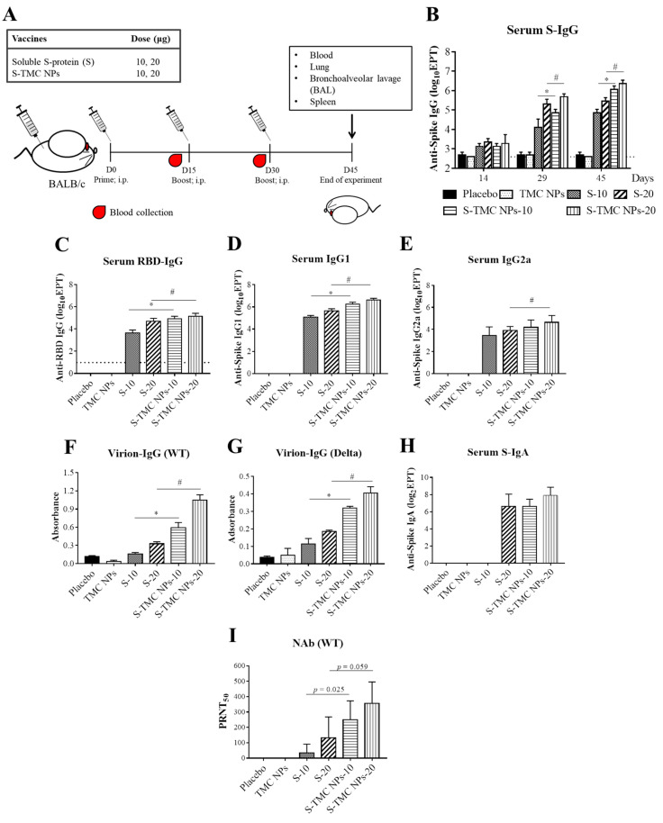
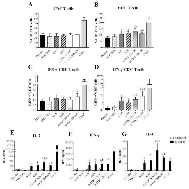
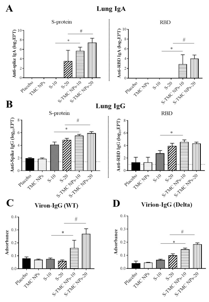
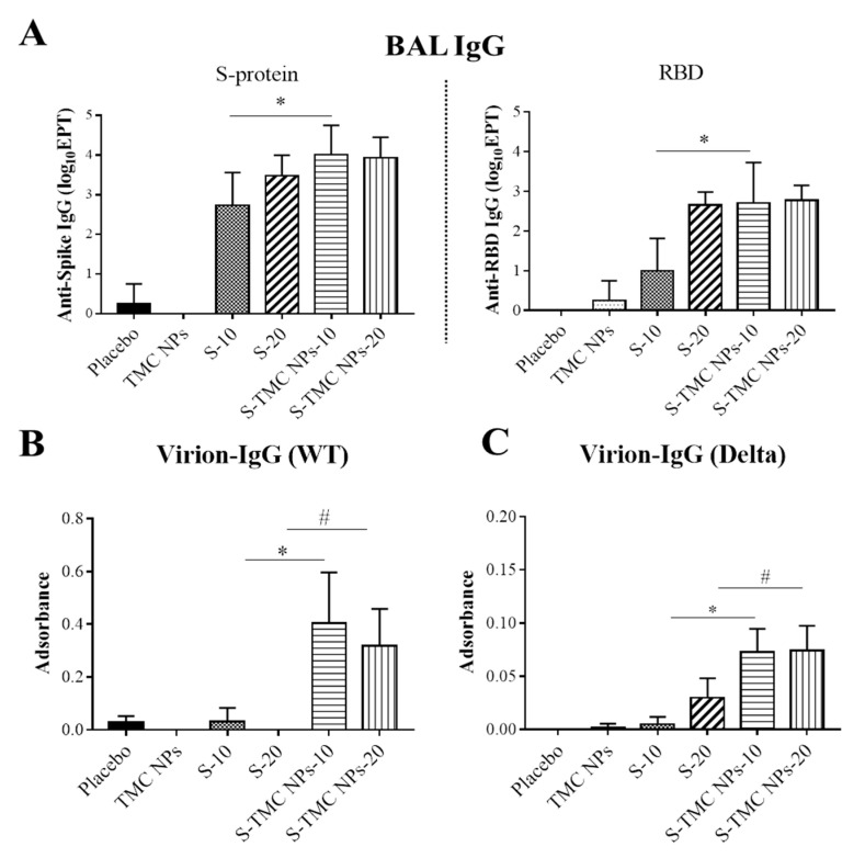
The COVID-19 pandemic has currently created an unprecedented threat to human society and global health. A rapid mass vaccination to create herd immunity against SARS-CoV-2 is a crucial measure to ease the spread of this disease. Here, we investigated the immunogenicity of a SARS-CoV-2 subunit vaccine candidate, a SARS-CoV-2 spike glycoprotein encapsulated in N,N,N-trimethyl chitosan particles or S-TMC NPs. Upon intraperitoneal immunization, S-TMC NP-immunized mice elicited a stronger systemic antibody response, with neutralizing capacity against SARS-CoV-2, than mice receiving the soluble form of S-glycoprotein. S-TMC NPs were able to stimulate the circulating IgG and IgA as found in SARS-CoV-2-infected patients. In addition, spike-specific T cell responses were drastically activated in S-TMC NP-immunized mice. Surprisingly, administration of S-TMC NPs via the intraperitoneal route also stimulated SARS-CoV-2-specific immune responses in the respiratory tract, which were demonstrated by the presence of high levels of SARS-CoV-2-specific IgG and IgA in the lung homogenates and bronchoalveolar lavages of the immunized mice. We found that peritoneal immunization with spike nanospheres stimulates both systemic and respiratory mucosal immunity.

Keywords: COVID-19 vaccine; SARS-CoV-2 spike glycoprotein; adjuvant delivery particles; mucosal immunity; peritoneal immunization.


Conflict of interest statement

The authors declare no conflict of interest.

Figures

Figure 1
Figure 1
The physical characterization of S-TMC NPs. The ionotropic gelation method was used to encapsulate SARS-CoV-2 spike proteins into TMC NPs. After NP preparation, the mean particle size and polydispersity index (PDI) of the S-TMC NPs were evaluated by a zetasizer (A). The entrapment of S-protein in TMC NPs was determined by Coomassie blue staining (B) and immunoblotting using the anti-His tag and anti-SARS-CoV-2 RBD antibodies (C). 1: Empty TMC NPs; 2: S-TMC NPs; 3: soluble S-antigen.
Figure 2
Figure 2
Cellular uptake of S-TMC NPs by phagocytic cells. Cultures of THP-1 cells were incubated with S-TMC NPs (20 μg/mL) or soluble S-protein (20 μg/mL) at 4 °C or 37 °C for 4 h. Intracellular S-antigens were detected by antibody staining with rabbit anti-SARS-CoV-2 antibody and analyzed by flow cytometry; (A) dot plot analysis, (B) the percentage of S-protein positive cells and (C) mean fluorescent intensity (MFI). The results are shown as means ± SD (n = 5); * indicates a significant difference between the soluble S-protein and the S-TMC NP treatment (p < 0.05).
Figure 3
Figure 3
Systemic humoral responses to soluble S-protein or S-TMC NPs. Mice were immunized intraperitoneally with three doses (2 weeks apart) of soluble S-protein (S) or S-TMC NPs (10 or 20 μg/dose). By Day 45 after immunization, samples from immunized mice, including blood, lungs, BALs and spleens, were harvested (A). Sera of immunized mice on Days 14, 29 and 45 were subjected to measurement of S-specific IgG by indirect ELISA (B). Sera on Day 45 were analyzed for RBD-specific IgG (C), S-specific IgG1 (D) and IgG2a (E) antibody ELISA assays. The levels of SARS-CoV-2-binding antibodies (Virion-IgG) in sera at a dilution of 1:100 were determined by capture ELISA against wild-type (WT) (F) and Delta variant viruses (G). IgA titers specific to purified S-protein were determined by indirect ELISA (H). The titers of serum neutralizing antibodies against SARS-CoV-2 were quantified by PRNT (I). Data are presented as means ± SD (n = 4); * and # indicate a significant difference between soluble S-protein and S-TMC NPs at 10 and 20 μg/dose, respectively (p < 0.05). Dotted line indicates the limit of detection (LOD) of the assay.
Figure 4
Figure 4
Cellular immune responses to soluble S-protein or S-TMC NPs. Splenocytes of immunized mice that received three doses of soluble S-protein or S-TMC NPs were isolated and stimulated ex vivo with 10 μg/mL of S-antigens for 72 h. Frequencies of S-specific CD4+ (A), CD8+ (B), IFN-γ+CD4+ (C) and IFN-γ+CD8+ (D) T cells in cultures of stimulated splenocytes were monitored by antibody staining, followed by analysis using flow cytometry. The levels of splenic cytokines, including IL-2 at 24 h (E), IFN-γ at 72 h (F) and IL-4 at 72 h (G) after antigen stimulation, were quantitated by ELISA. The results are presented as means ± SD (n = 4); “a” indicates significant differences between placebo and soluble S-protein or S-TMC NPs. “b” indicates significant differences between TMC NPs and S-TMC NPs. “c and d” indicate significant differences between soluble S-protein and S-TMC NPs at 10 and 20 μg/dose, respectively (p < 0.05).
Figure 5
Figure 5
Mucosal responses to soluble S-protein or S-TMC NPs in the lungs of immunized mice. Lungs of immunized mice were collected on Day 45 and processed by homogenization. The levels of S- and RBD-specific IgA (A) or IgG (B) antibodies in lung homogenates were detected by indirect ELISA. Binding antibodies against SARS-CoV-2 particles (virion–IgG) of WT (C) and Delta variant viruses (D) present in the lung homogenates diluted at 1:100 was determined by capture ELISA. Data are means ± SD (n = 4); * and # indicate a significant difference between soluble S-protein and S-TMC NPs at 10 and 20 μg/dose, respectively (p < 0.05). Dotted line indicates the LOD of the assay.
Figure 6
Figure 6
Mucosal responses to soluble S-protein or S-TMC NPs in the BALs of immunized mice. BALs were harvested from immunized mice on Day 45 and subjected to quantitation of anti-S and anti-RBD IgG antibodies using indirect ELISA (A). BALs diluted at 1:10 were analyzed for virion–IgG capture ELISA assays against WT SARS-CoV-2 (B) and the Delta variant strain (C). The results are shown as means ± SD (n = 4); * and # indicate a significant difference between the mice immunized with soluble S-protein and S-TMC NPs at 10 and 20 μg/dose, respectively (p < 0.05).

Similar articles

Cited by

References

    1. Tabibzadeh A., Esghaei M., Soltani S., Yousefi P., Taherizadeh M., Tameshkel F.S., Golahdooz M., Panahi M., Ajdarkosh H., Zamani F., et al. Evolutionary study of COVID-19, severe acute respiratory syndrome coronavirus 2 (SARS-CoV-2) as an emerging coronavirus: Phylogenetic analysis and literature review. Vet. Med. Sci. 2021;7:559–571. doi: 10.1002/vms3.394. - DOI - PMC - PubMed
    1. Rastogi M., Pandey N., Shukla A., Singh S.K. SARS coronavirus 2: From genome to infectome. Respir. Res. 2020;21:318. doi: 10.1186/s12931-020-01581-z. - DOI - PMC - PubMed
    1. Kasuga Y., Zhu B., Jang K.J., Yoo J.S. Innate immune sensing of coronavirus and viral evasion strategies. Exp. Mol. Med. 2021;53:723–736. doi: 10.1038/s12276-021-00602-1. - DOI - PMC - PubMed
    1. de Assis R.R., Jain A., Nakajima R., Jasinskas A., Felgner J., Obiero J.M., Norris P.J., Stone M., Simmons G., Bagri A., et al. Analysis of SARS-CoV-2 antibodies in COVID-19 convalescent blood using a coronavirus antigen microarray. Nat. Commun. 2021;12:6. doi: 10.1038/s41467-020-20095-2. - DOI - PMC - PubMed
    1. Piccoli L., Park Y.J., Tortorici M.A., Czudnochowski N., Walls A.C., Beltramello M., Silacci-Fregni C., Pinto D., Rosen L.E., Bowen J.E., et al. Mapping Neutralizing and Immunodominant Sites on the SARS-CoV-2 Spike Receptor-Binding Domain by Structure-Guided High-Resolution Serology. Cell. 2020;183:1024–1042.e21. doi: 10.1016/j.cell.2020.09.037. - DOI - PMC - PubMed

Publication types

MeSH terms