Review
. 2021 Nov 4;13(11):2218.
doi: 10.3390/v13112218.

Stephan Oroszlan and the Proteolytic Processing of Retroviral Proteins: Following A Pro

Affiliations
Review

Stephan Oroszlan and the Proteolytic Processing of Retroviral Proteins: Following A Pro

Ronald Swanstrom et al. Viruses. .

Abstract

Steve Oroszlan determined the sequences at the ends of virion proteins for a number of different retroviruses. This work led to the insight that the amino-terminal amino acid of the mature viral CA protein is always proline. In this remembrance, we review Steve's work that led to this insight and show how that insight was a necessary precursor to the work we have done in the subsequent years exploring the cleavage rate determinants of viral protease processing sites and the multiple roles the amino-terminal proline of CA plays after protease cleavage liberates it from its position in a protease processing site.

Keywords: HIV-1; Oroszlan; capsid; protease; retroviruses.


Conflict of interest statement

The authors declare no conflict of interest.

Figures
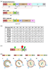
Figure 1
Figure 1
The Gag Polyprotein Precursor, Its Protease Cleavage Sites, and Proteolytic Processing During Virion Maturation. (a) The simplest organization of the retroviral Gag, Pro, and Pol coding domains consisting of MA (matrix), CA (capsid), NC (nucleocapsid), PR (protease), RT-RH (reverse transcriptase with its associated RNase H activity), IN (integrase), and the subunits of the Env protein SU (surface) and TM (transmembrane). The red line indicates the RNA splicing event that creates the subgenomic mRNA needed to express the env gene. The Gag protein and the gag gene have separate writing conventions to designate the protein versus the gene. The HIV-1 Gag protein has two additional spacer peptides (SP1 and SP2), as well as the “late domain” p6 polypeptide, which is involved in virion budding. The Gag-Pro-Pol precursor is formed by a frameshifting event at the NC/SP1 boundary that reads the p6 domain in an alternative reading frame (Transframe/TF), which is in frame with the pro and pol reading frames. (b) The major protease cleavage site sequences in the HIV-1 Gag and Gag-Pro-Pol precursors. The cleavage site sequences are written N to C, with the cleavage occurring between P1 and P1’. These cleavage site sequences are highly conserved as individual sequences even though they vary significantly between cleavage sites. The P1’ prolines are bolded. (c) The cleavage sites in HIV-1 Gag are shown, with the large red arrow indicating the first cleavage at the SP1/NC junction, the green arrows show the next cleavages between MA/CA and SP2/p6, and the small blue arrows show the slow removal of the two spacer peptides. The resulting processing intermediates are shown below from left to right. (d) Intact Gag and an immature virus particle are shown on the left, then the sequential cleavage intermediates and changes in virion protein arrangement are shown until a mature virion is formed.
Figure 2
Figure 2
Proteolytic Processing at the HIV-1 MA-CA Junction Refolds the HIV-1 CA N-terminus into a β-hairpin Conformation that Buries the N-terminal CA Pro 1 Residue. (a) Proteolytic processing (scissors) of the immature Gag polyprotein at the MA–CA junction of the Gag1-278 polyprotein (blue, PDB: 2GOL) creates a new CA N-terminus and drives folding of the N-terminal CA β-hairpin (dashed circle, green, PDB: 2GON). (b) The mature N-terminal CA Pro1 (P1) fits snugly into a pocket in which the buried, protonated N-terminus makes a salt bridge with Asp51 and a hydrogen bond with Gln13 (blue dashed lines), and Pro1 Cδ makes hydrophobic packing interactions with Ala14 and Ile15 (black dashed lines, PDB: 4B4N).
Figure 3
Figure 3
The HIV-1 CA β-hairpin Adopts Different Conformations that Open and Close the Central CA Hexamer Pore. (a) Space-filling model showing the central pore of the CA hexamer in its closed (green, PDB: 5TSX) and open (magenta, PDB: 5HGL) configurations. (b) Positions of the β-hairpin (middle panel) in the closed (green, PDB: 4B4N) and open (magenta, PDB: 5HGL) conformations. Note the rotation about CA Pro1 (P1, sticks). Expanded panels show the Pro1 polar interactions (dashed orange lines) and surrounding CA residues in the closed (green, left panel, PDB: 2GON) and open (magenta, right panel, PDB: 5HGL) conformations. The red sphere depicts a water molecule.
Figure 4
Figure 4
HIV-1 CA Residue R18 Forms a Basic Collar that Binds Translocating Nucleotide Triphosphates and IP6. (a) Cross-section of the mature CA hexamer bound to dATP, highlighting how the basic R18 “arginine collar” residues (sticks) interact with the γ- and β-phosphates of a translocating nucleotide triphosphate (PDB: 5HGM). Dashed lines depict the poorly structured loop of the β-hairpins in the closed conformation. Note that for clarity, we are showing only one of the six rotationally disordered dATP ligands. (b) Cross-section of the mature CA hexamer highlighting how IP6 can bind above and below the arginine collar (PDB: 6BHT). Note that for clarity, we are showing only one of each of the six rotationally disordered IP6 ligands.
Figure 5
Figure 5
Relative Rates of Protease Cleavage of SP1/NC and MA/CA Cleavage Sites with Substitutions. The wild-type sequence is shown in bold and underlined (MA/CA: SQNY/PIVQ; SP1/NC: ATIM/MQRG). The substitutions are shown above or below in single-letter amino acid code. The change in the rate of cleavage relative to wild type is shown by the vertical positioning of the indicated substitution, with faster rates above the wild-type sequence and slower rates below, as indicated by the Y-axis label. Changes within three-fold represent little to no change. The P2 and P2’ positions are indicated by the red rectangles. The red double-headed arrow notes the change in specificity in P2 and P2’. Data are reproduced from ref [79].
Figure 6
Figure 6
Structural Interpretation of Substrate Orientation Within the HIV-1 Protease with and without a P1’ Proline. The surfaces of the viral protease are shown for the S2 and the S2’ subsites with yellow as hydrophobic, blue as positive charge, and red as negative charge. Two substrates are superimposed in stick figure with MA/CA in orange and SP1/NC in green. The individual substrate amino acids are indicated, with arrows showing the differing orientations of the P2 and P2’ side chains in the substrate. The structures are from ref. [80], and the figure was constructed in PyMol using PDB files 1KJ4 and 1KJ7.

References

    1. Oroszlan S.I., Rizvi S., O’Connor T.E., Mora P.T. Use of Synthetic Polyglucose for Density-Gradient Centrifugation of Viruses. Nat. Cell Biol. 1964;202:780–781. doi: 10.1038/202780a0. - DOI - PubMed
    1. Oroszlan S., Johns L.W., Rich M.A. Ultracentrifugation of a murine leukemia virus in polymer density gradients. Virology. 1965;26:638–645. doi: 10.1016/0042-6822(65)90327-2. - DOI - PubMed
    1. Gilden R.V., Lee Y.K., Oroszlan S., Walker J.L., Huebner R.J. Reptilian C-type virus: Biophysical, biological, and immunological properties. Virology. 1970;41:187–190. doi: 10.1016/0042-6822(70)90069-3. - DOI - PubMed
    1. Oroszlan S., Fisher C.L., Stanley T.B., Gilden R.V. Proteins of the Murine C-Type RNA Tumour Viruses: Isolation of a Group-specific Antigen by Isoelectric Focusing. J. Gen. Virol. 1970;8:1–10. doi: 10.1099/0022-1317-8-1-1. - DOI - PubMed
    1. Oroszlan S., Foreman C., Kelloff G., Gilden R.V. The group-specific antigen and other structural proteins of hamster and mouse C-type viruses. Virology. 1971;43:665–674. doi: 10.1016/0042-6822(71)90290-X. - DOI - PubMed

Personal name as subject

LinkOut - more resources