

COVID-19 Anosmia: High Prevalence, Plural Neuropathogenic Mechanisms, and Scarce Neurotropism of SARS-CoV-2?
- PMID: 34835030
- PMCID: PMC8625547
- DOI: 10.3390/v13112225
COVID-19 Anosmia: High Prevalence, Plural Neuropathogenic Mechanisms, and Scarce Neurotropism of SARS-CoV-2?
Abstract
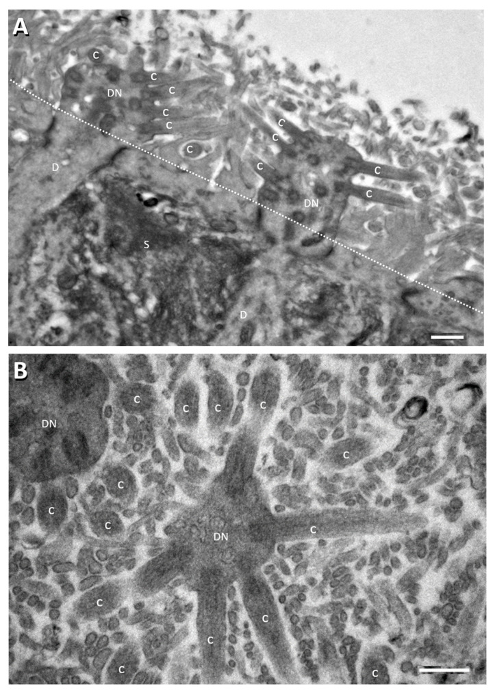
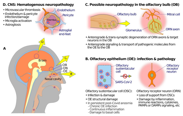
Severe acute respiratory syndrome coronavirus 2 (SARS-CoV-2) is the causative pathogen of coronavirus disease 2019 (COVID-19). It is known as a respiratory virus, but SARS-CoV-2 appears equally, or even more, infectious for the olfactory epithelium (OE) than for the respiratory epithelium in the nasal cavity. In light of the small area of the OE relative to the respiratory epithelium, the high prevalence of olfactory dysfunctions (ODs) in COVID-19 has been bewildering and has attracted much attention. This review aims to first examine the cytological and molecular biological characteristics of the OE, especially the microvillous apical surfaces of sustentacular cells and the abundant SARS-CoV-2 receptor molecules thereof, that may underlie the high susceptibility of this neuroepithelium to SARS-CoV-2 infection and damages. The possibility of SARS-CoV-2 neurotropism, or the lack of it, is then analyzed with regard to the expression of the receptor (angiotensin-converting enzyme 2) or priming protease (transmembrane serine protease 2), and cellular targets of infection. Neuropathology of COVID-19 in the OE, olfactory bulb, and other related neural structures are also reviewed. Toward the end, we present our perspectives regarding possible mechanisms of SARS-CoV-2 neuropathogenesis and ODs, in the absence of substantial viral infection of neurons. Plausible causes for persistent ODs in some COVID-19 convalescents are also examined.
Keywords: COVID-19; SARS-CoV-2; anosmia; olfactory dysfunction; pathogenesis.
Conflict of interest statement
The authors declare no conflict of interest.
Figures
Similar articles
-
Receptors Involved in COVID-19-Related Anosmia: An Update on the Pathophysiology and the Mechanistic Aspects.Int J Mol Sci. 2024 Aug 5;25(15):8527. doi: 10.3390/ijms25158527. Int J Mol Sci. 2024. PMID: 39126095 Free PMC article. Review.
-
Evidence of SARS-CoV2 Entry Protein ACE2 in the Human Nose and Olfactory Bulb.Cells Tissues Organs. 2020;209(4-6):155-164. doi: 10.1159/000513040. Epub 2021 Jan 22. Cells Tissues Organs. 2020. PMID: 33486479 Free PMC article.
-
Regeneration Profiles of Olfactory Epithelium after SARS-CoV-2 Infection in Golden Syrian Hamsters.ACS Chem Neurosci. 2021 Feb 17;12(4):589-595. doi: 10.1021/acschemneuro.0c00649. Epub 2021 Feb 1. ACS Chem Neurosci. 2021. PMID: 33522795 Free PMC article.
-
Massive transient damage of the olfactory epithelium associated with infection of sustentacular cells by SARS-CoV-2 in golden Syrian hamsters.Brain Behav Immun. 2020 Oct;89:579-586. doi: 10.1016/j.bbi.2020.06.032. Epub 2020 Jul 3. Brain Behav Immun. 2020. PMID: 32629042 Free PMC article.
-
The Molecular Basis of Olfactory Dysfunction in COVID-19 and Long COVID.Lifestyle Genom. 2024;17(1):42-56. doi: 10.1159/000539292. Epub 2024 May 15. Lifestyle Genom. 2024. PMID: 38749402 Review.
Cited by
-
Clinical factors influencing olfactory performance in patients with persistent COVID-19 smell loss longer than 1 year.Laryngoscope Investig Otolaryngol. 2023 Oct 9;8(6):1449-1458. doi: 10.1002/lio2.1160. eCollection 2023 Dec. Laryngoscope Investig Otolaryngol. 2023. PMID: 38130252 Free PMC article.
-
Type 2 and Non-type 2 Inflammation in the Upper Airways: Cellular and Molecular Alterations in Olfactory Neuroepithelium Cell Populations.Curr Allergy Asthma Rep. 2024 Apr;24(4):211-219. doi: 10.1007/s11882-024-01137-x. Epub 2024 Mar 16. Curr Allergy Asthma Rep. 2024. PMID: 38492160 Free PMC article. Review.
-
Olfactory dysfunction in COVID-19: new insights into the underlying mechanisms.Trends Neurosci. 2023 Jan;46(1):75-90. doi: 10.1016/j.tins.2022.11.003. Epub 2022 Nov 16. Trends Neurosci. 2023. PMID: 36470705 Free PMC article. Review.
-
[Headache associated with COVID-19 vaccination: how to classify?].Schmerz. 2023 Jun;37(3):185-194. doi: 10.1007/s00482-022-00687-1. Epub 2023 Jan 16. Schmerz. 2023. PMID: 36645522 Free PMC article. German.
-
Spontaneous recovery of anosmia after 2.5 years in a young COVID-19 patient.Eur Clin Respir J. 2023 Feb 13;10(1):2178598. doi: 10.1080/20018525.2023.2178598. eCollection 2023. Eur Clin Respir J. 2023. PMID: 36815941 Free PMC article. No abstract available.
References
Publication types
MeSH terms
Substances
Grants and funding
LinkOut - more resources
Full Text Sources
Medical
Miscellaneous
